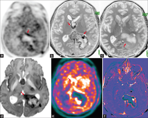
Figure 3.
Operated case of anaplastic astrocytoma (World Health Organization grade III, isocitrate dehydrogenase1 (IDH1) negative, not otherwise specified (NOS) with recurrence (red arrows) shows peripheral rim of hypermetabolic lesions (18F-fluoroethyl-L-tyrosine positron emission tomography AC (a) and color-coded positron emission tomography images (e) in septum pellucidum, splenium of the corpus callosum, left parieto-occipital lobes. There is peripheral rim enhancement (b) with washout in 75 min delayed contrast images (c), corresponding marginal diffusion restriction (d) and contrast rim enhancement in the subtracted T1 contrast images (f)

