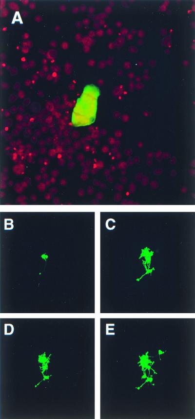
FIG. 1.
Cell-to-cell spread of MVeGFP in undifferentiated and differentiated murine neuroblastoma cells. TMN cells at a confluency of 80% were infected with MVeGFP at an MOI of 0.01. Undifferentiated TMN cells were fixed and examined by CSLM for fluorescence. (A) Syncytium of infected undifferentiated TMN cells at 90 h.p.i. Nuclei were counterstained using propidium iodide, and micrographs represent a 10-μm composite optical section. Infected dTMN cells were identified by UV microscopy, and the positions of infectious centers were marked to aid in their reidentification throughout a time period of 42 h. (B) Two infected dTMN cells, one of which has an extended process (48 h.p.i.). (C) A number of newly infected cells with interconnecting autofluorescent processes (66 h.p.i.). (D) No additional fluorescent cells were observed by 72 h.p.i., but a number of additional autofluorescent processes were visible. By 90 h.p.i., further TMN cells became infected via these processes (E). Autofluorescent images (B to E) were collected as single optical sections by CSLM.

