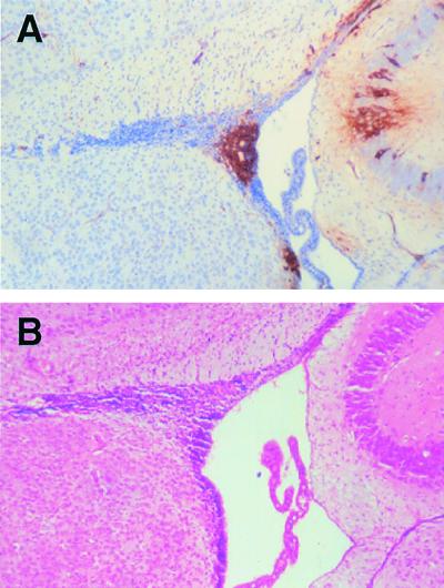
FIG. 4.
Immunohistochemistry and H&E staining of brain sections from Ifnarko-CD46Ge suckling animals infected intracerebrally with the rodent brain-adapted virus CAM/RB. Sections were formalin fixed, and MV antigen was detected with an antinucleocapsid monoclonal antibody; positive staining appears brown. (A) Virus-infected neuroblasts and ependymal cells surrounding the ventricle. (B) H&E staining was used to confirm the identify of the infected cells. Magnification, ×100.

