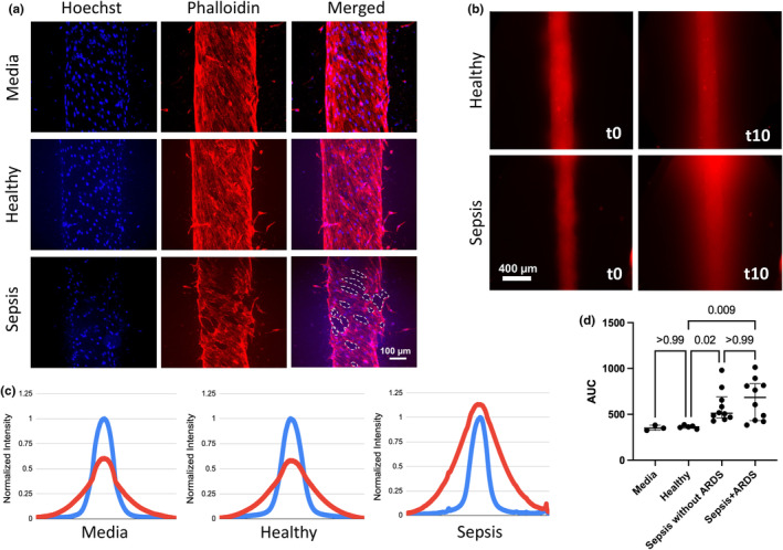
FIGURE 1.

Lung endothelial MPS incubated with cell culture media (“media”), healthy donor plasma (“healthy”), and sepsis patient plasma (“sepsis”) for 16 h. (a) Staining of lumens incubated with media, healthy and sepsis plasma. Dashed white lines indicate holes in EC coverage in sepsis lumen. (b) Fluorescent dextran imaged just after addition to lumens (“t0”) and 10 min later (“t10”). (c) Plots of fluorescence intensity across lumens incubated with media, healthy and sepsis plasma. Intensity normalized to minimum (0) and maximum (1) at t0 (blue line) and t10 (red line). (d) Increased vascular permeability in lumens incubated with sepsis + ARDS and sepsis without ARDS plasma (n = 10 Sepsis+ARDS plasma, median 684.9, IQR 365.7; n = 10 Sepsis without ARDS plasma, median 511.9, IQR 164.6) compared with healthy plasma (n = 5 healthy controls, median 363.1, IQR 14.9, p = 0.009 for difference with Sepsis+ARDS plasma, p = 0.02 for difference with Sepsis without ARDS plasma) or media (median 349.5 IQR 27.9.)
