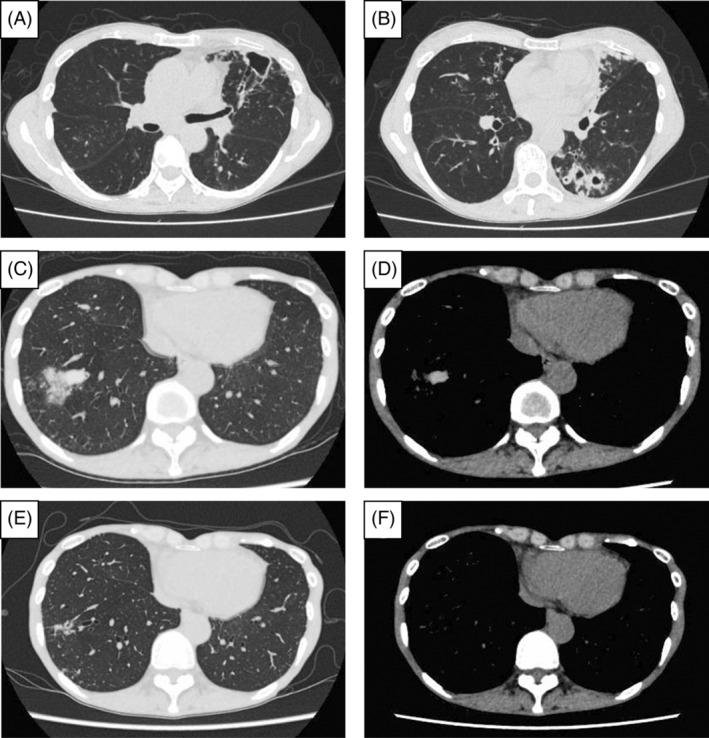
FIGURE 1.

Imaging findings of chest CT at the diagnosis of NTM‐PD, at the diagnosis of ABPA, and after the initiation of dupilumab. CT showed cavities with their wall thickness and bronchiectasis in the upper and lower lobe of the left lung (A, B). At the diagnosis of ABPA, CT showed a high attenuation mucus plug in the lower lobe of the right lung (C, D). After initiating dupilumab, mucus plugging improved, and bronchiectasis was observed in the same lesion (E, F). ABPA, allergic bronchopulmonary aspergillosis; CT, computed tomography; NTM‐PD, nontuberculous mycobacterial‐pulmonary disease.
