Fig. 1.
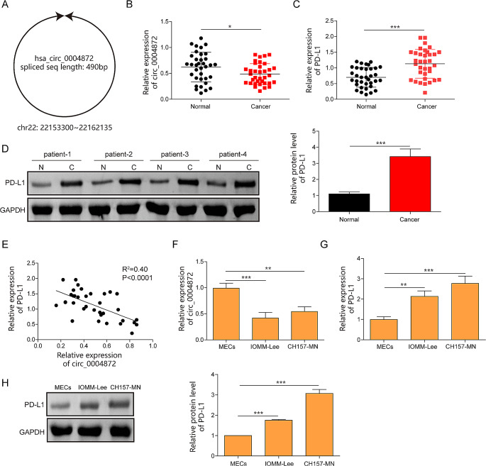
Hsa_circ_0004872 was downregulated and PD-L1 was upregulated in meningiomas. A The schematic diagram of the hsa_circ_0004872. B The expression of hsa_circ_0004872 in meningiomas tissues was determined by RT-qPCR (n = 35). C The mRNA expression of PD-L1 in meningiomas tissues was evaluated by RT-qPCR (n = 35). D The protein expression of PD-L1 was tested by western blot (n = 35). E The correlation between hsa_circ_0004872 and PD-L1 analyzed by Pearson correlation analysis. F, G The expression of hsa_circ_0004872 and PD-L1 were determined by RT-qPCR in meningiomas cells (IOMM‐ Lee, CH157-MN) and MEC cells. H The protein of PDL1 in meningiomas cells (IOMM‐ Lee, CH157-MN) and MEC cells was quantified by western blot. *P < 0.05. **P < 0.01

