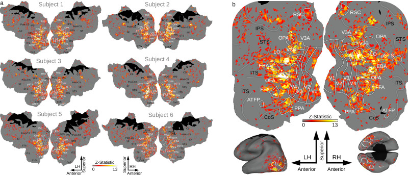
Fig. 2. CSVA model voxel-wise prediction accuracy scores mapped onto cortex.
a Voxels where activity was significantly predicted by the Combined Semantic, Valence and Arousal (CSVA) model are shown on cortical maps for all 6 subjects. For each voxel, prediction accuracies were calculated using the z-transformed correlation between the CSVA model predicted time-course and the recorded BOLD time-course for the validation dataset. Significance was assessed by permutation testing, see Methods for details. The CSVA model significantly predicts validation BOLD time-courses across much of OTC. This was consistently observed across subjects. b Prediction accuracy scores for subject 1; the cortical map is cropped (top) to zoom in on OTC. In addition, prediction accuracy scores are projected onto inflated lateral (bottom left) and ventral (bottom right) cortical surfaces. Note: Regions of interest (ROIs) are labeled in white, sulci in black. RSC Retrosplenial Complex, OPA Occipital Place Area, LO Lateral Occipital cortex, pSTS Posterior Superior Temporal Sulcus, EBA Extrastriate Body Area, OFA Occipital Face Area, FFA Fusiform Face Area, PPA Parahippocampal Place Area, ATFP Anterior Temporal Face Patch. IPS Intraparietal Sulcus, STS Superior Temporal Sulcus, ITS Inferior Temporal Sulcus, CoS Collateral Sulcus.

