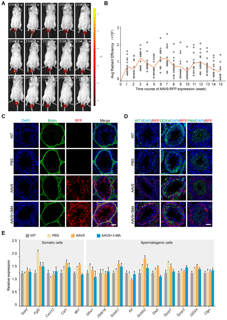
Figure 2.
In vivo kinetics and safety assessment of the co-injection of AAV9 and 3-MA into the testis. (A) Representative sequential fluorescence imaging of the RFP signal (in mouse 210#) at indicated time-points (weeks 1-15) after co-injection of AAV9-CMV-RFP+3-MA, determined using an in vivo imaging system (IVIS). Dark red and yellow indicate low and high RFP expression, respectively, following AAV9-CMV-RFP administration. (B) Kinetics of AAV9-CMV-RFP expression in the mouse testis in vivo at the indicated time-points. Average (Avg) radiant efficiency was determined from the fluorescence imaging results. Sequential fluorescence imaging data were generated from each mouse at each time-point (n = 15). (C) Results of the biotin tracer experiment to determine BTB integrity. The mouse testes (at PND 21) were first microinjected with PBS, AAV9-CMV-RFP, or AAV9-CMV-RFP+3-MA and then interstitially injected with biotin (green). Samples were recovered 30 min after biotin microinjection (n = 3). Scale bar: 50 μm. (D) Immunostaining of testicular sections for WT1 (green, left), DDX4 (green, middle), and PNA (green, right) from testes microinjected with PBS, AAV9-CMV-RFP, or AAV9-CMV-RFP+3-MA. Nuclei were stained with DAPI (blue). Scale bar: 50 μm. (E) qRT-PCR analysis of representative functional gene expression in somatic and spermatogenic cells isolated from testes microinjected with PBS, AAV9-CMV-RFP, or AAV9-CMV-RFP+3-MA. Data were normalized to Actb expression and are presented as the mean ± SEM (n = 3).

