Fig. 4.
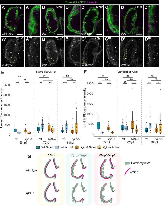
Llgl1 promotes timely deposition of laminin to the apical ventricular surface. (A-D‴) Confocal z-slices of the ventricle of Tg(myl7:LifeAct-GFP) transgenic embryos visualising the myocardium (green) and anti-laminin antibody (magenta) in wild-type siblings (A,C) and llgl1 mutant embryos (B,D). Yellow boxed areas are enlarged to the right. A-D and A″-D″ show a merge view, A′-D′ and A‴-D‴ show laminin staining. llgl1 mutants still exhibit basal laminin at 72 hpf (B), and are yet to clearly establish apical laminin at 80 hpf (D). Scale bar=25 µm. (E) Quantification of apicobasal laminin intensity in the outer curvature shows that laminin is basally enriched in both wild-type siblings and llgl1 mutants at 55 hpf (wild-type, n=27; llgl1 mutants, n=26). Laminin is apically enriched in wild-type siblings from 72 hpf onwards (72 hpf, n=48; 80 hpf, n=49), whereas llgl1 mutants exhibit no apical laminin enrichment at 72 hpf (n=40), and only apical enrichment at 80 hpf (n=68). (F) Quantification of apicobasal laminin intensity in the ventricular apex reveals that both wild-type siblings and llgl1 mutants have enrichment of basal laminin at 55 hpf (wild type, n=25; llgl1 mutants, n=23). Wild-type siblings do not exhibit significant degradation of basal laminin and enrichment of apical laminin until 80 hpf (72 hpf, n=45; 80 hpf, n=42), whereas llgl1 mutants still retain significant levels of basal laminin at 72 hpf (n=40), and do not have significant apical deposition even by 80 hpf. (E,F) Kruskal–Wallis test with multiple comparisons (n=58) (****P<0.0001, ***P<0.001, **P<0.01, *P<0.05). ns, non significant. Box plots show median values (middle bars) and first to third interquartile ranges (boxes); whiskers indicate 1.5× the interquartile ranges; dots indicate data points. (G) Schematic depicting dynamics of laminin distribution in wild type and llgl1 mutants.

