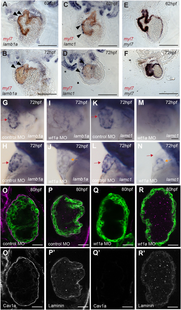
Fig. 5.
The epicardium deposits laminin onto the apical ventricular surface. (A-F) Sections through the hearts of two-colour mRNA in situ hybridisation analysis of lamb1a (A,B), lamc1 (C,D) and myl7 controls (E,F) in blue, combined with myl7 expression (red) to highlight the myocardium, at 62 hpf and 72 hpf (n=3 per gene/stage). lamb1a and lamc1 are expressed in epicardial cells adjacent to/on the surface of the myocardium (arrowheads), contrasting with the myl7 blue/red control demonstrating colocalised myocardial stains. Scale bars: 200 μm. A-F show coronal sections. (G-N) mRNA in situ hybridisation analysis of lamb1a and lamc1 expression in control MO and wt1a MO-injected embryos at 72 hpf. G,I,K,M show ventral views, H,J,L,N show lateral views. Scale bars: 25 µm. Control MO-injected embryos express lamb1a and lamc1 around the ventricle (red arrow, lamc1 n=6, lamb1a n=5), whereas wt1a MO-injected embryos lose expression of both lamb1a and lamc1a around the ventricle (lamc1 n=9, lamb1a n=6) but retain low levels in the atrium (orange arrow). (O-R′) Confocal z-slices of the ventricle of 80 hpf Tg(myl7:HRAS-GFP) transgenic embryos injected with either control MO (O,P) or wt1a MO (Q,R), and an anti-Cav1a antibody (magenta; O,Q) or anti-laminin antibody (magenta; P,R). Scale bars: 25 µm. Control MO-injected embryos exhibit expression of Cav1a (n=9/10) and laminin (n=10/10) at the apical CM surface (O,P), whereas wt1a MO-injected embryos show loss of both Cav1a (Q, n=9/9) and laminin (R, n=10/10) expression. O-R show a merge view, O′-R′ show Cav1a or laminin staining.

