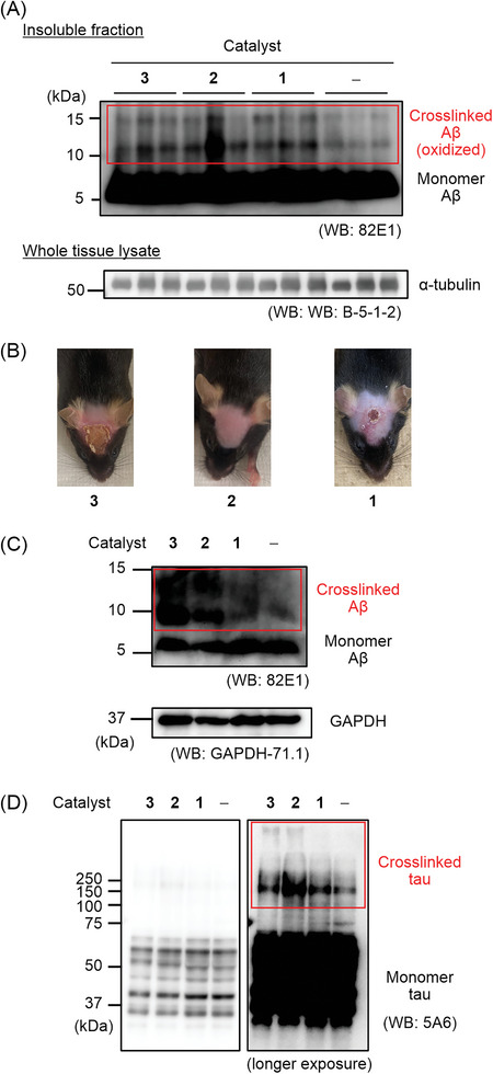
Figure 6.

In vivo and ex‐vivo photooxygenation reaction. A) In vivo photooxygenation reaction. A solution of EV 3, LEV 2, or ABB 1 was intravenously injected into 5–6 month old AppNL‐G‐F/NL‐G‐F mice (n = 3 experiments, each group) expressing human Arctic Aβ. After an interval, the mice were irradiated with LED (λ = 595 nm) for 10 min. The operation set (catalyst injection and photoirradiation) was repeated five times over 5 d. At 24 h after the final operation set, the brain was excised and homogenized using a 1× PBS buffer. After the fractionation, the insoluble fraction was analyzed by SDS‐PAGE using a 15% Tris‐Tricine gel and Western blot (WB) using an anti‐Aβ antibody. For loading controls, α‐tubulin in lysates before fractionation was analyzed. B) Photos of mouse scalp after the photooxygenation treatment in (A). C, D) Catalytic photooxygenation of human brain lysate. The temporal cortex of an AD patient was homogenized using a 10× volume of PBS (containing cOmplete EDTA+ (Roche) and PhosSTOP (Sigma)). A catalyst (2.5 × 10⁻⁶ m) was added to the brain lysate and the mixture was irradiated with 595 nm light for 3 h or kept in the dark at 37 °C. The resulting mixture was analyzed with SDS‐PAGE and WB (anti‐Aβ antibodies: 82E1 (IBL) and anti‐GAPDH antibodies: GAPDH‐71.1 for (C), anti‐tau antibodies: 5A6 for (D)). Human AD‐tau is comprised of 6 isoforms, whose sizes are 36.8–45.9 kDa. CBB staining was shown in Figure S30 (Supporting Information).
