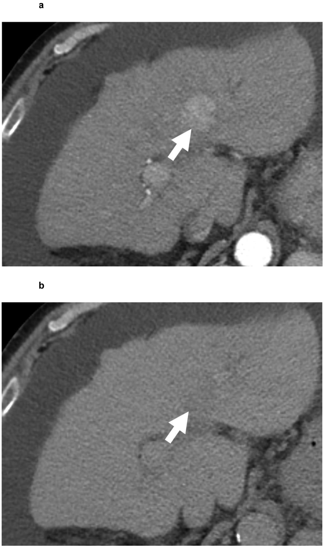
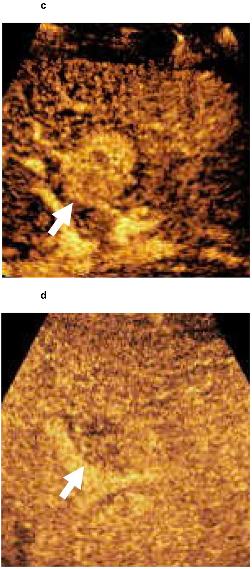
Figure 4:


67-year-old patient with a 3.0 cm segment 2 LI-RADS 4 observation (white arrow) showing (a) APHE and (b) questionable washout in multiphase CT examination. Given the risks associated with liver biopsy in the patient, contrast-enhanced ultrasound confirmed a LI-RADS 5 segment 2 observation with (c) APHE and (d) faint washout. HCC diagnosis by CEUS is not approved for liver transplantation purposes. However, as the patient was not considered for liver transplantation because of non-HCC factors, a biopsy was not performed.
