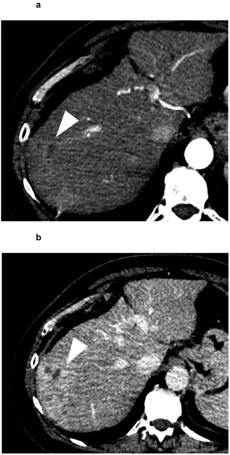
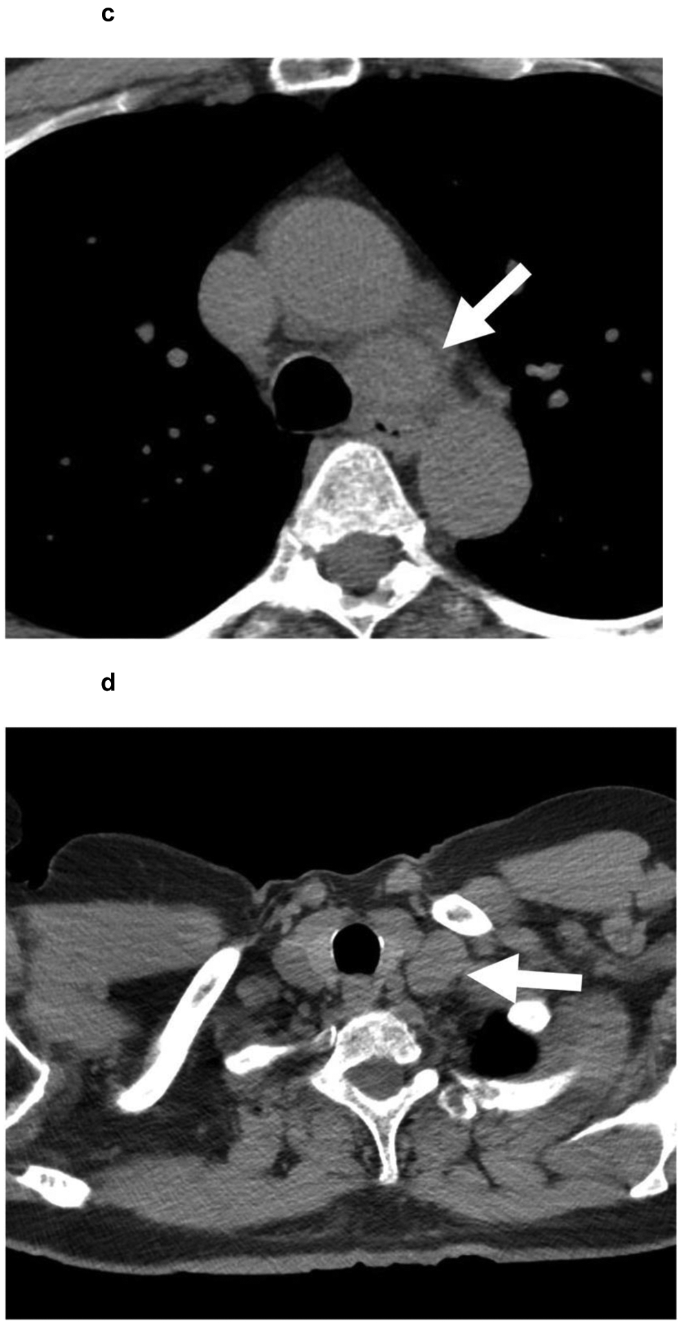
Figure 5:


A 62-year-old patient with (a,b) a 7.4 cm segment 7 infiltrative LR-M observation (white arrowhead) showing (a) APHE and (b) persistent delayed heterogeneous enhancement. Biopsy revealed HCC. (c,d) Noncontrast CT of the chest reveals enlarged (c) mediastinal and (d) supraclavicular lymph nodes (white arrows). The patient was referred to systemic therapy.
