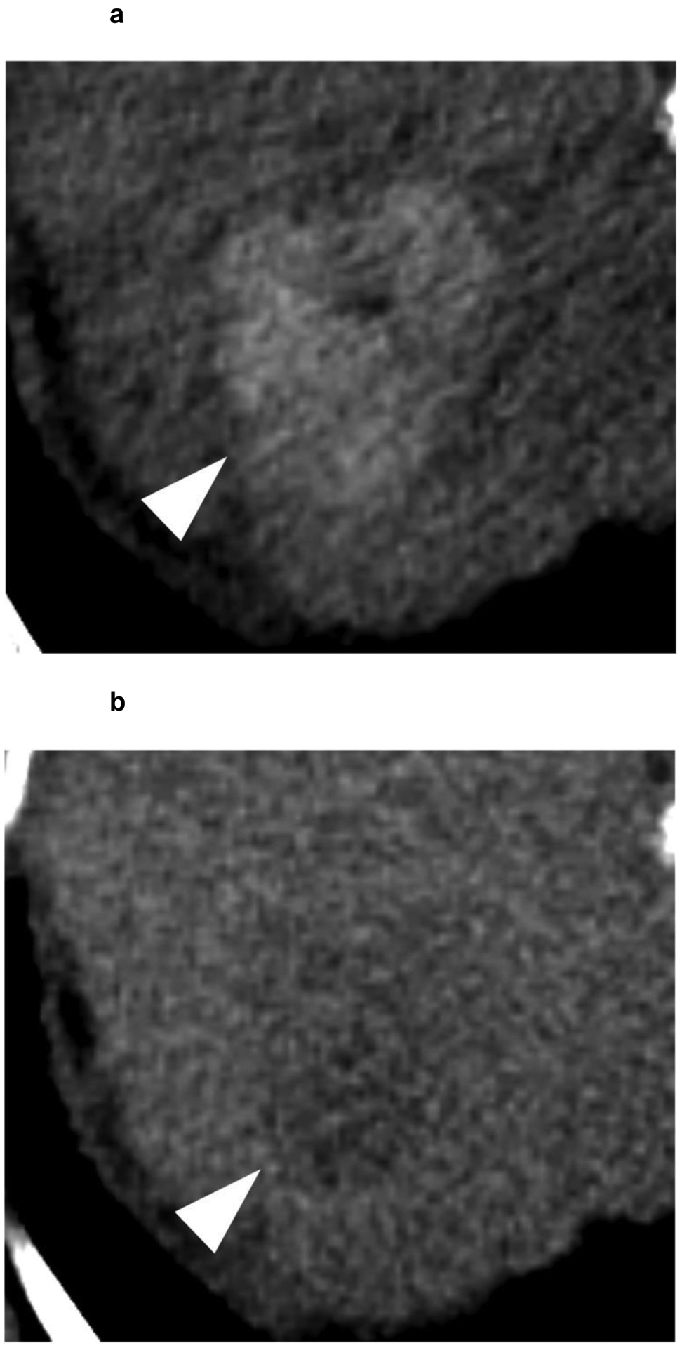
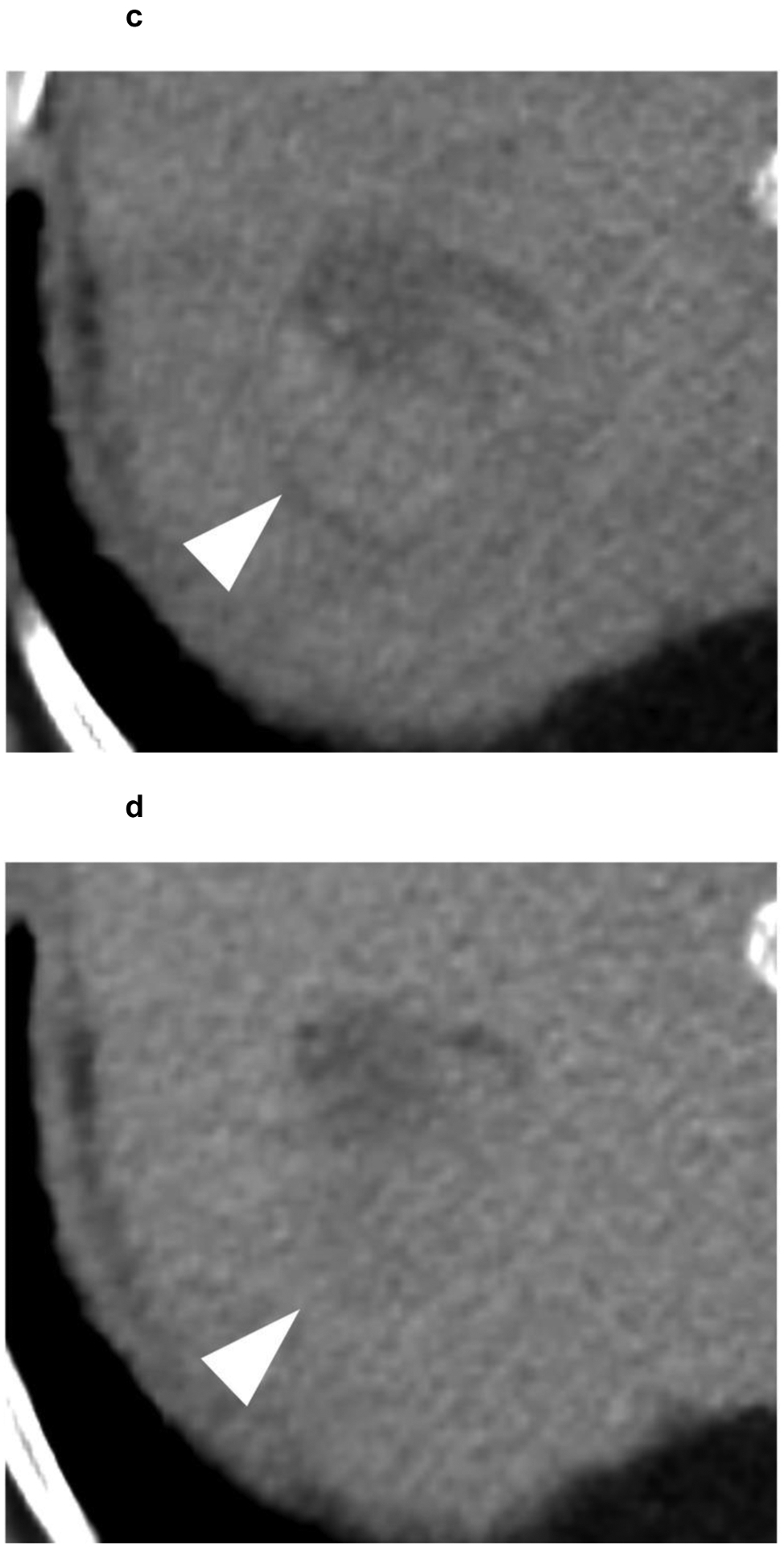
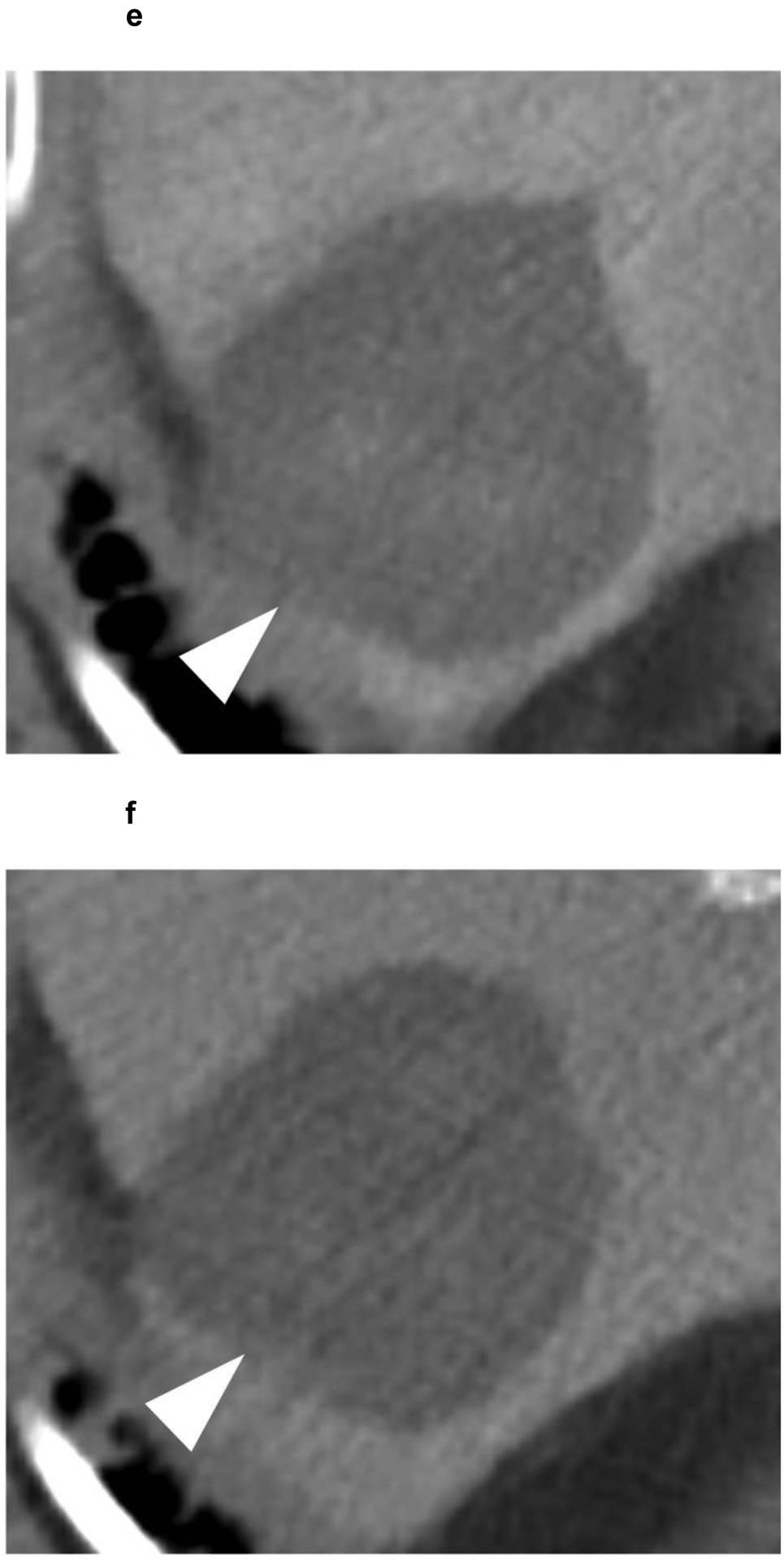
Figure 6:


A 63-year-old patient presents with a 4.1 cm segment 7 LI-RADS 5 observation (white arrowhead) with (a) APHE and (b) washout appearance. A CT performed 3 months after SBRT shows (c) residual enhancement with (d) equivocal washout (LI-RADS TR equivocal). (e,f) The arterial and delayed phases from CT performed 7 months after SBRT reveal resolution of the suspicious enhancement (LI-RADS TR non-viable). No intervening additional treatment was performed after the SBRT or during the follow-up period.
