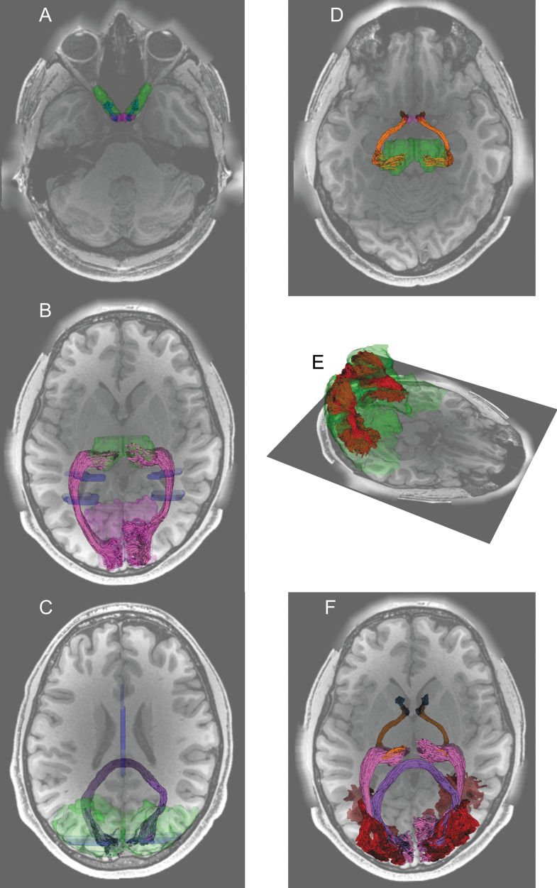
Fig. 2.
Visual white matter tracts were identified by tractography in dMRI data from representative participants in the Human Connectome Project Young Adult data.250 A: The optic nerve (blue) was identified by tractography and mask ROIs (green) generated by automated segmentation of a structural image.106 The optic nerve and the optic chiasm (purple) were overlaid on an axial section of a T1-weighted image. B. The optic radiation (magenta) was identified using waypoint ROIs (blue) and endpoint ROIs (thalamus, green; primary visual cortex, light purple) transformed from the Montreal Neurological Institute (MNI) template space.182 C. The forceps major (dark purple) was identified from a waypoint ROI (blue) in the corpus callosum and endpoint ROIs (green) in the occipital cortex of each hemisphere. D. The optic tract (orange) was identified by using the optic chiasm (purple) and the thalamus (green) identified by segmentation on structural images as endpoint ROIs. E. The vertical occipital fasciculus (red) was identified by using dorsal and ventral visual areas (green) identified by using the automated anatomical labeling atlas.276 F. All identified tracts were overlaid on an axial section of a T1-weighted image.

