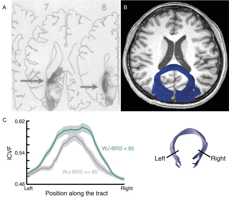
Fig. 5.
Forceps major. A: The lesion topography of alexia patients.191 The dark gray area (highlighted by arrow) indicates the brain lesion site commonly appeared in alexia patients. This area corresponds to the forceps major. Reprinted by permission from reference 191. B: The forceps major (blue) identified by using tractography in dMRI data overlaid on the axial section of a T1-weighted image. C: Tractometry study on the forceps major. Left panel: Tract profile of the forceps major in good (dark green) and poor readers (gray).202 The horizontal axis depicts the normalized position along the forceps major, whereas the vertical axis depicts microstructural measurement (ICVF estimated by NODDI).34 The shaded area indicates ±1 s.e.m. The reading performance was measured by the WJ-BRS. Right panel: the forceps major identified by tractography. Reprinted by permission from reference 202 (under the CC BY-NC-ND license). dMRI, diffusion-weighted MRI; ICVF, intra-cellular volume fraction; NODDI, neurite orientation dispersion and density imaging; WJ-BRS, Woodcock-Johnson Basic Reading Score.

