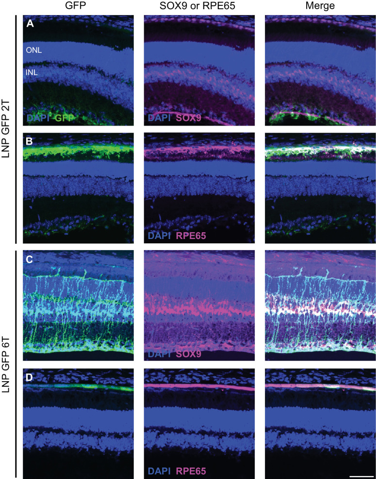
Figure 3.
LNP delivery of EGFP mRNA transfects Müller glia and RPE cells in the mouse retina. Retinal cross-sections were co-stained with DAPI, anti-GFP, and either anti-SOX9 or anti-RPE65 immunofluorescence 48 hours after intravitreal injections of EGFP mRNA encapsulated in 2Tor 6T LNPs. (A) For the 2T formulation, cells labelled with anti-GFP colocalize with nuclei labelled with anti-SOX9 in the inner nuclear layer, indicating EGFP expression in the Müller glia. (B) Cells that express EGFP also express RPE65, indicating EGFP expression in the RPE. Similarly, for 6T, there is co-expression of EGFP and (C) SOX9 and (D) RPE65. Images shown are representative of at least three retinas (scale bar = 50 µm).

