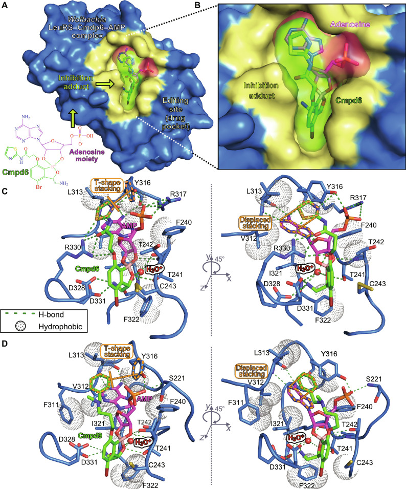
Fig. 3. Crystal structures of the Wolbachia LeuRS complexes with AMP-compound adducts.
(A) Overall structure of the Wolbachia LeuRS complex with the adduct AMP-Cmpd6 bound into the editing site. Van der Waals surface representation in blue was used for the protein, with the drug-binding site in yellow. (B) Zoomed-in view showing the inhibition adduct in surface and stick representations, with the AMP group in pink and Cmpd6 in green. (C and D) Main interactions established by the adenosine-drug inhibition adducts formed by Cmpd6 and Cmpd9, respectively. Hydrogen bonds are shown as green dashed lines and hydrophobic interactions are shown as dotted spheres. Key protein residues are shown as blue sticks, the hydroxonium ion is shown as a red sphere, and the compound-AMP adducts are in the same color code as in (B). π-π aromatic stackings of the thiazole/phenyl rings of Cmpd6/9 are highlighted in orange dashed lines. For clarity, panels are rotated by 45° around the y axis.

