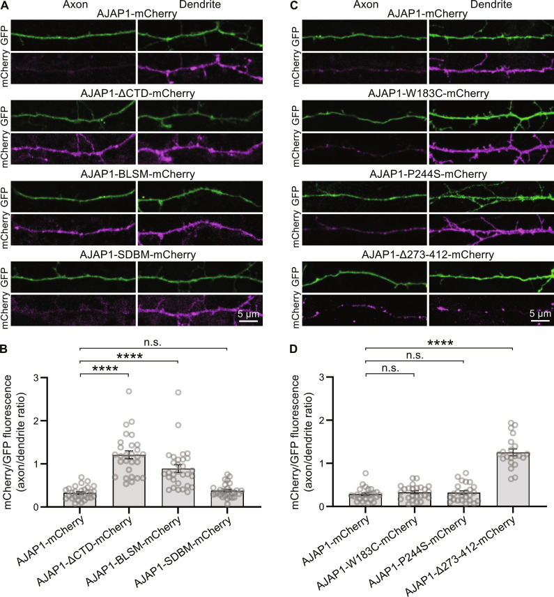
Fig. 3. Intracellular sorting signals target AJAP1 to the dendrites.
(A) Axonal versus dendritic sorting of AJAP1-mCherry fusion constructs expressed in cultured hippocampal neurons. AJAP1-mCherry constructs were coexpressed with the volume marker GFP at DIV 7 and fluorescence intensities quantified after 4 days. AJAP1-mCherry and AJAP1-SDBM-mCherry exhibit a dendritic localization. AJAP1-ΔCTD-mCherry and AJAP1-BLSM-mCherry lacking BLSS are distributed to axons and dendrites. (B) Ratios of mCherry fluorescence normalized to GFP between axons and dendrites for constructs shown in (A). ****P < 0.0001, n.s., P > 0.05, Kruskal-Wallis and Dunn’s multiple comparisons test, n = 28 to 30 cells per condition. (C) Subcellular sorting of AJAP1 variants expressed in cultured hippocampal neurons. AJAP1-W183C-mCherry and AJAP1-P244S-mCherry exhibit a dendritic localization. AJAP1-∆273–412-mCherry lacking BLSS is distributed to axons and dendrites. (D) Ratios of mCherry fluorescence normalized to GFP between axons and dendrites for constructs shown in (C). n.s., P > 0.05, ****P < 0.0001, Kruskal-Wallis test and Dunn’s multiple comparisons test, n = 21 to 24 cells per condition.

