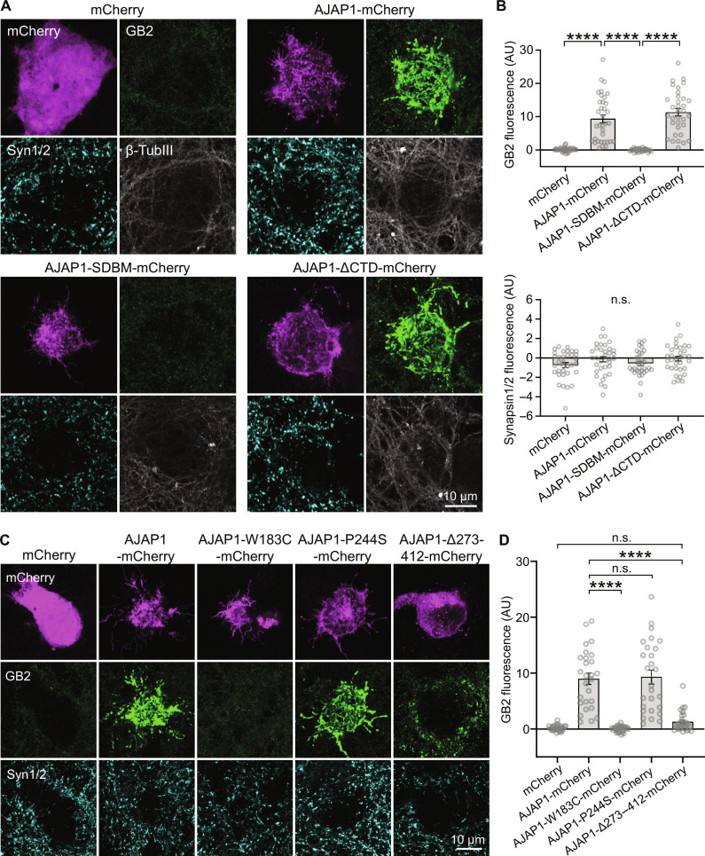
Fig. 5. Recruitment of neuronal GBRs by AJAP1 variants in trans.
(A) Transcellular recruitment of neuronal GBRs to HEK293T cells expressing AJAP1 constructs. HEK293T cells expressing AJAP1-mCherry, AJAP1-SDBM-mCherry or AJAP1-ΔCTD-mCherry were cocultured for 36 hours with hippocampal neurons (DIV12). Immunolabeling for GB2 shows a strong transcellular recruitment of GBRs to the soma of HEK293T cells expressing AJAP1-mCherry and AJAP1-ΔCTD-mCherry but not AJAP1-SDBM-mCherry. AJAP1-expressing cells did not recruit Synapsin1/2. β-TubulinIII was used as a marker for neurites. (B) GB2 and Synapsin1/2 immunofluorescence at the soma of HEK293T cells expressing AJAP1 constructs. The background fluorescence in areas devoid of transfected HEK293T cells was subtracted. n.s., P > 0.05, ****P < 0.0001, Welch’s ANOVA, Dunnett’s T3 multiple comparisons test (GB2), Kruskal-Wallis test and Dunn’s multiple comparisons test (Synapsin1/2), n = 33 to 35 cells per condition. (C) Transcellular recruitment of neuronal GBRs to HEK293T cells expressing AJAP1-mCherry and the variants AJAP1-W183C-mCherry, AJAP1-P244S-mCherry, and AJAP1-Δ273–412-mCherry. (D) GB2 immunofluorescence at the soma of HEK293T cells expressing AJAP1 variants. n.s., P > 0.05, ****P < 0.0001, Kruskal-Wallis test and Dunn’s multiple comparisons test, n = 27 to 28 cells per condition.

