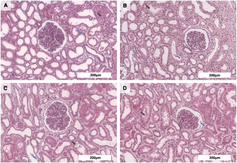
Figure 3.
Histological analyses of kidneys after normothermic EVKP using OCs. Representative images of hematoxylin and eosin stained renal cortex regions: Kidneys perfused with no OCs (A), blood (B), PFC-based OCs (C), or M101 (D) showed necrotic cell detritus within the tubular lumen (blue arrow) as well as a mild tubular vacuolization (black arrow), indicating a potentially reversible mild to moderate acute tubular injury and minor signs of tubular necrosis. However, overall renal morphology of the individual groups was intact (scale bar: 200 µm).

