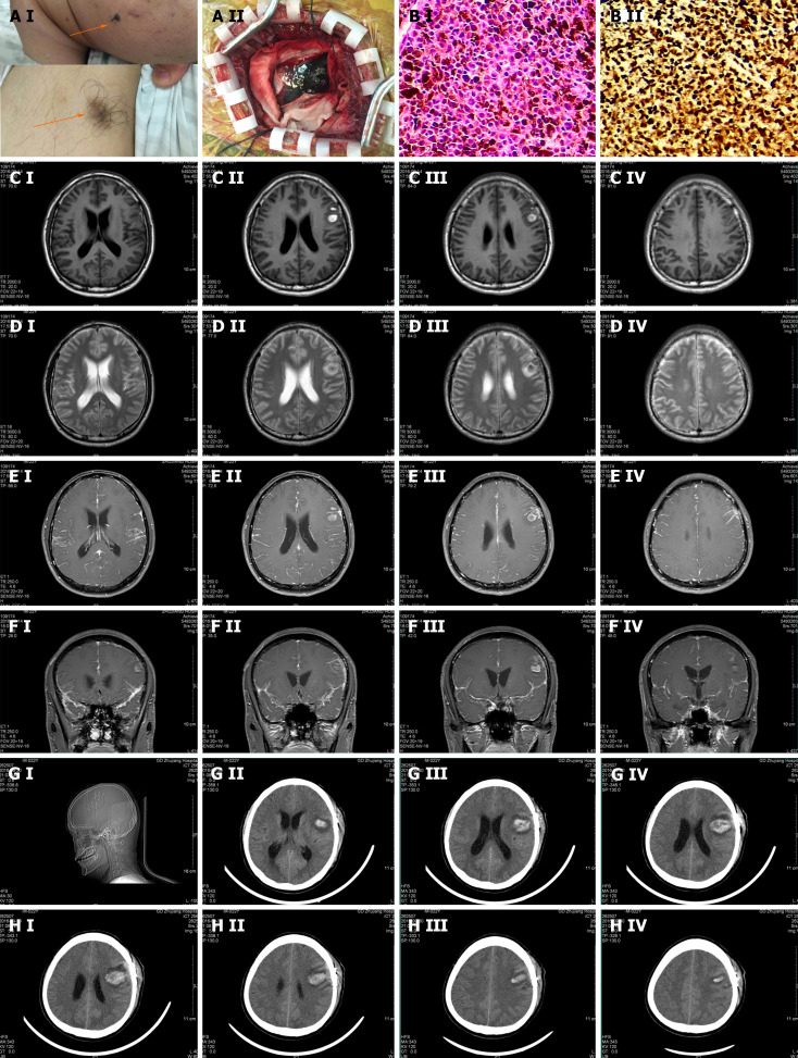
Figure 1.
Pathological and imaging features of our melanoma patient. A: A-I: On the left cheek, there was a hairy nevus with the size of 1.5 cm × 2.2 cm. The color was relatively dark, and the edge blurred. It was as hard as a nasal tip and there was no ulcer around. Meanwhile, there was a hairy pigment spot on the inside left lower arm with the size of 3.1 cm × 2.5 cm. Its color was relatively light and the edge blurred. The rigidity was the same as the former. A-II: Histopathological examination: (Left frontal lobe lesion) the tumor tissue appears nest-like, with tumor cells showing polygonal shape, abundant cytoplasm, significant melanin content, large and deeply stained nuclei with irregular features. Immunohistochemistry: VIM (+), HMB45 (+), s-100 (+), Melin-A (+), P53 (+), CK (-), 70% Ki-67 (+). Pathological diagnosis: (Malignant melanoma) of the left frontal lobe. B: B-I: The distribution of carcinoma cells showed as nests (200 ×). The tumor cells were polygonal. The cell plasma was rich with a large amount of melanin in it. The nucleus was large and irregular. B-II: The tumor cells in immunohistochemical staining showed: VIM (+), HMB45 (+), S-100 (+), Melin-A (+), P53 (+), CK (-), KI-67 70% (+). CEDF: MRI showed a patch of long T1 and T2 signals on the left frontal lobe with nodular short T1 and T2 signals inside, and it was not enhanced in the enhancement scanning. However, in the enhancement scanning, the meninges were widely thickening and enhanced, especially around the abnormal signal lesion on the left frontal lobe and the tentorium of the cerebellum. There was a poor contrast agent filling in the superior sagittal sinus; C-I, II, III, and IV: T1 scan of the focal area; D-I, II, III, and IV: T2 scan of the focal area; E-I, II, III, and IV: Enhancement scan of the focal area; F-I, II, III, and IV: Enhancement scan on the focal area in coronal position; G and H: The head computed tomography (CT) scan after showed that there was still a high-density shadow on the left frontal lobe and the range got larger, inside new high-density shadow with the range of 23 mm × 45 mm was detected and the CT attenuation value was 58HU, and there was an edema zone around. In the adjacent sulci, there was a line-like high-density shadow. The ventricular system was enlarged.

