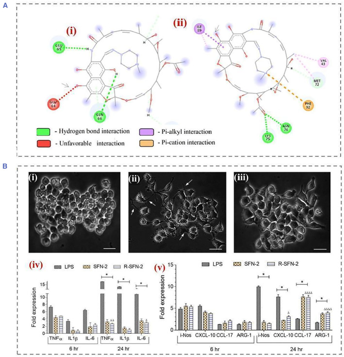
FIGURE 8.

(A) Interaction of (i) RIF with silk fibroin at pH 7.2; arrows indicate unfavorable electrostatic interactions between the 4‐hydroxyl group of rifampicin and GLU64 of silk fibroin (red). (ii) RIF and silk fibroin at pH interactions at 3.8; arrows indicate the formation of favorable interactions. (B) Effects of pH‐induced self‐assembled silk fibroin nanoparticles (SFN‐2, 100 μg/mL) on the morphology of RAW 264.7 cells observed by phase contrast microscopy after 24 h. (i) Untreated naive macrophages, (ii) lipopolysaccharide‐treated macrophages, and (iii) R‐SFN‐2‐treated macrophages. (iv) Proinflammatory cytokine expression profile of RAW264.7 macrophages when exposed to SFN‐2. (v) Anti‐inflammatory cytokine expression profiles upon exposure to SFN2. Reproduced with permission. 44 Copyright 2022, American Chemical Society.
