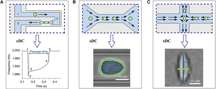
FIGURE 2.

The schematic operation principles of three types of microfluidic technologies for cell deformability cytometry, including constriction deformability cytometry (A), fluid shear deformability cytometry (B), and extensional flow deformability cytometry (C). The bottom rows represent the typical signals of each method. Reproduced under terms of the CC‐BY license. 10 Copyright 2020, The Authors, published by Springer Nature.
