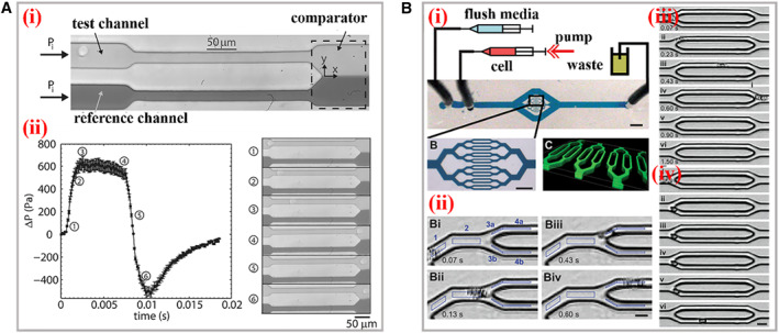
FIGURE 4.

Practical applications of cDCs. (A) (i) The structure and working mechanism of the microfluidic cell squeezer. A balanced interface in the comparator region between the fluids in the reference and test channels. (ii) The curve between excess pressure drop and time. The optical images of different states of the interface within the squeezer corresponding to the cellular deformations. Reproduced with permission. 21 Copyright 2013, AIP Publishing. (B) (i) The scheme of the biophysical flow cytometer device. (ii) This visual tracking of neutrophil. The images of neutrophil transiting through microchannels before (iii) and after (iv) exposure to the inflammatory mediator increases. Reproduced with permission. 24 Copyright 2008, The Royal Society of Chemistry.
