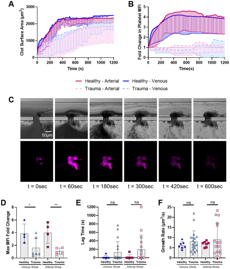
Fig 2. Despite similar amounts of clot deposited, trauma patient samples have massively reduced platelet contribution to microfluidic clot.

A Trauma patient samples had slightly reduced clot surface area deposition but overall deposited substantial amount of clot at the injury site. B Trauma patient samples had massively reduced platelet deposition compared to healthy controls at both venous and arterial shear. C Representative trauma microfluidic hemostasis timelapse from a sample that achieved hemostasis. Transmitted light (top) and platelet (anti-CD41) fluorescence (bottom, pink). D Maximum platelet MFI fold change was significantly reduced in trauma patient samples. Traces are shown as mean lines with standard deviation indicated as error bars below. E, F Lag time and growth rate of total surface area deposition were not different between trauma and controls. Individual data points are shown with bars at mean and error bars indicating standard deviation. ns: not significant * p<0.05, ** p<0.01, *** p<0.001. MFI: mean fluorescence intensity.
