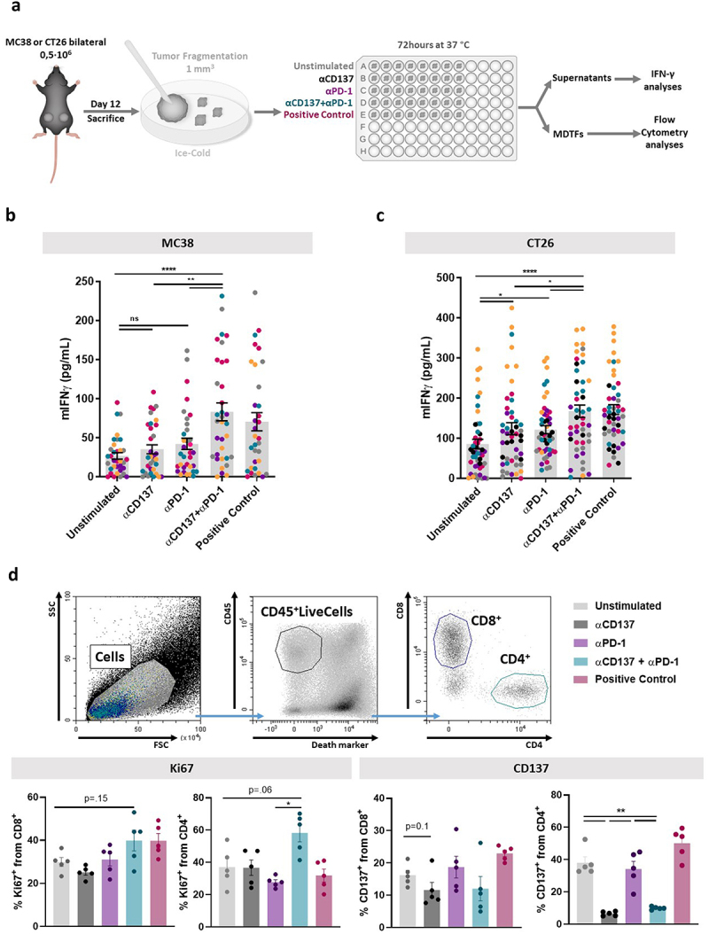
Figure 1.

Tumor-infiltrating lymphocytes in cultured tumor fragments from established mouse tumors are activated by anti-PD-1 plus anti-CD137 mAb combinations.
a) Experimental schematic representation of tumor fragment cultures in 96× well plates, seeding one fragment per well and culturing for 72 h to retrieve supernatants and prepare cell suspensions. b and c) IFNγ concentrations in the supernatants following cultures of MC38 (b) and CT26 (c) fragments stimulated with the indicated antibodies or concanavalin A as a positive control. Dots represent single wells and colors individual mice. d) Flow cytometry dot-plots showing a representative case of MC38-derived cell suspensions in which live CD4+ and CD8+ cells can be observed and electronically gated by FACS (upper panels). Lower panels show percentages of Ki67+ CD8+ and CD4+ cells and percentages of CD137+ cells by surface staining from the different conditions tested. For these flow cytometry experiments, cell suspensions from the fragments were pooled and dots represent cultures from individual mice bearing bilateral tumors. MDTFs: mouse-derived tumor fragments.
