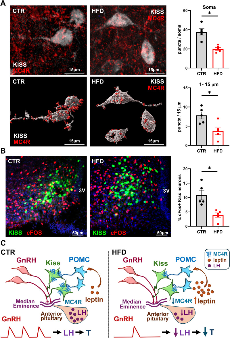
Figure 7.
Fewer active kisspeptin neurons in HFD mice compared with CTR. A, Kiss-EGFP (n = 5) mice were fed CTR or HFD for 12 weeks, after which immunohistochemistry was performed to analyze MC4R and cFOS levels. Top, MC4R receptor (red) colocalized to kisspeptin neurons, identified by GFP (white); Bottom, 3D reconstruction; Right, MC4R puncta in 15–25 neurons per mouse were counted and averaged. Each dot represents one animal, bars represent group average, * indicates statistical difference between CTR and HFD by t test (top, p = 0.0014; bottom, p = 0.0162). B, Left, Images of Kiss-GFP and cFOS after CTR and HFD; Right, percent of cFOS positive kisspeptin neurons determined through colocalization of cFOS (red) and kisspeptin neurons (green). Each dot represents one animal, bars represent group average, * indicates statistical difference between CTR and HFD by t test (p = 0.0052). C, Model of HFD-mediated effects on hypothalamic circuitry, based on results presented here. Left, GnRH secretion into the median eminence capillaries is regulated by kisspeptin neurons that integrate input from POMC neurons. Pulsatile secretion of GnRH stimulates LH synthesis and secretion from the anterior pituitary, which in turn regulates testosterone synthesis in the gonads. Right, HFD-induced obesity leads to increased leptin that causes higher activity of POMC neurons and downregulation of MC4R, a receptor for POMC product αMSH, in kisspeptin neurons. HFD exposure results in lower activity of kisspeptin neurons, which leads to lower pulse frequency of GnRH secretion, and diminished levels of LH and T in the circulation.

