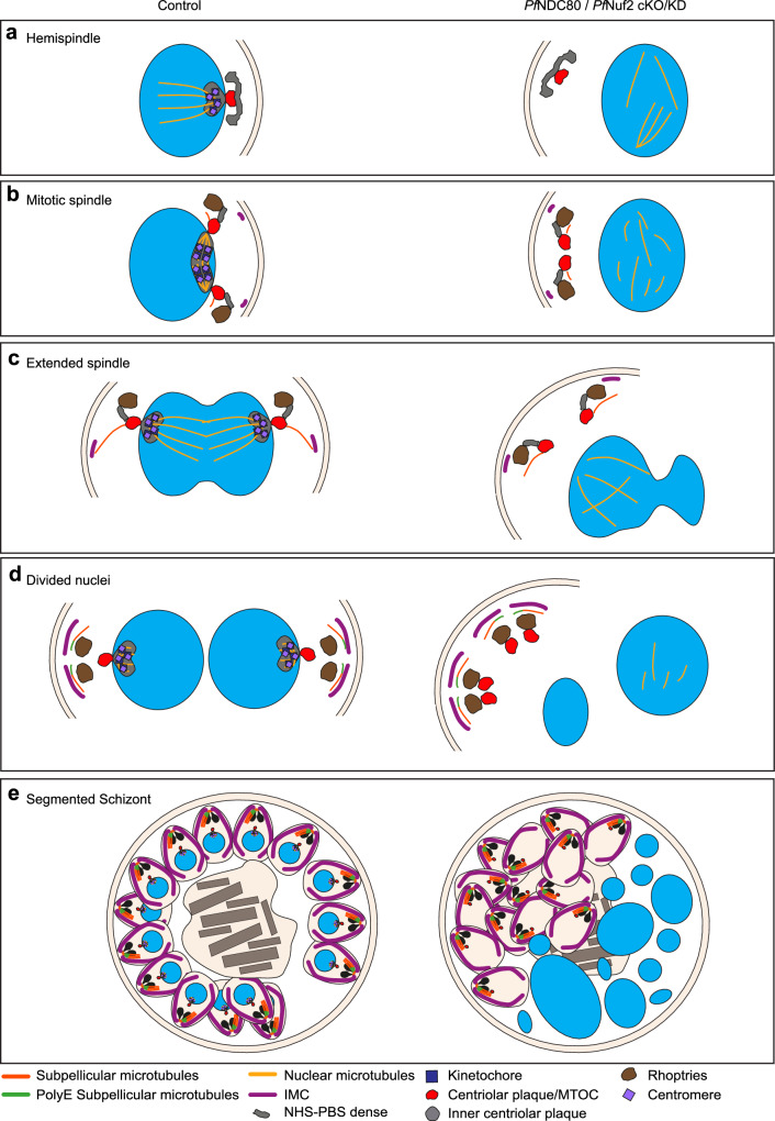
Fig. 6. Parasite spindle structures with and without kinetochore associated proteins.
Diagram illustrating the apparent locations of different parasite structures during mitosis; and disruption of organisation upon loss of the kinetochore components. (a) Hemispindle. (b) Mitotic spindle. (c) Extended spindle. (d) Divided nuclei. (e) Segmented schizont. In kinetochore-disrupted parasites, the mitotic spindle is disorganised, the inner and outer centriolar plaque structures are separated and the nexus between the mitotic machinery and the apical complex is lost.

