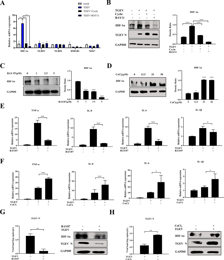
Fig 6.
HIF-1α positively regulates TGEV-induced inflammation downstream of the RIG-I–NF-κB pathway. (A and B) Apical-out organoids were infected with TGEV followed by Cyclo (1 mM) or BAY11 (2 µM) treatment for 48 h. Subsequently, transcription levels of HIF-1α, NLRP3, NLRP6, HMGB1, and NEK7 in the apical-out organoids post TGEV infection were measured by RT-qPCR (A) and HIF-1α protein expression was determined by Western blotting and analyzed using ImageJ (B). (C and D) Apical-out organoids were treated with the indicated concentrations of BAY87 or CoCl2 for 48 h, then HIF-1α protein expression was detected by Western blotting and analyzed by ImageJ. (E and F) Apical-out organoids were infected with TGEV followed by BAY87 (5 µM) or CoCl2 (25 µM) treatment for 48 h, then transcription levels of TNF-α, IL-8, IL-6, and IL-1β were determined by RT-qPCR. (G and H) Apical-out organoids were infected with TGEV followed by BAY87 (5 µM) or CoCl2 (25 µM) treatment for 48 h, then TGEV viral load was detected by RT-qPCR, and TGEV N and HIF-1α protein expressions were measured by Western blotting. Results are presented as mean ± SD of data from three independent experiments *, P ≤ 0.05; **, P ≤ 0.01; ***, P ≤ 0.001, determined by two-tailed Student’s t test.

