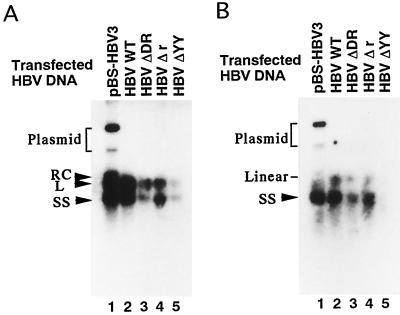
FIG. 4.
Southern blot analysis of HBV DNA in viral or core particles. (A) HBV DNA in viral particles. HBV particles secreted into the culture medium of the cells transfected with pBS-HBV3, WT HBV, HBV ΔDR, HBV Δr, or HBV ΔYY (lanes 1 to 5) were treated with 1 mg of proteinase K per ml and 1% SDS and then directly subjected to 1% agarose gel electrophoresis. The resultant DNA was blotted to the filter paper and hybridized with an HBV DNA probe. Arrowheads indicate the positions corresponding to three different forms of HBV DNA (RC, L, and SS) and the bracket shows the position of transfected plasmid DNA. (B) HBV DNA in core particles was treated as described for panel A. Lanes 1 to 5 contain the samples from pBS-HBV3-, WT-HBV-, HBV ΔDR-, HBV Δr-, and HBV ΔYY-transfected cells, respectively. The arrowhead indicates the position of the SS form of HBV DNA. The positions of the transfected plasmid and linear DNA are also indicated.

