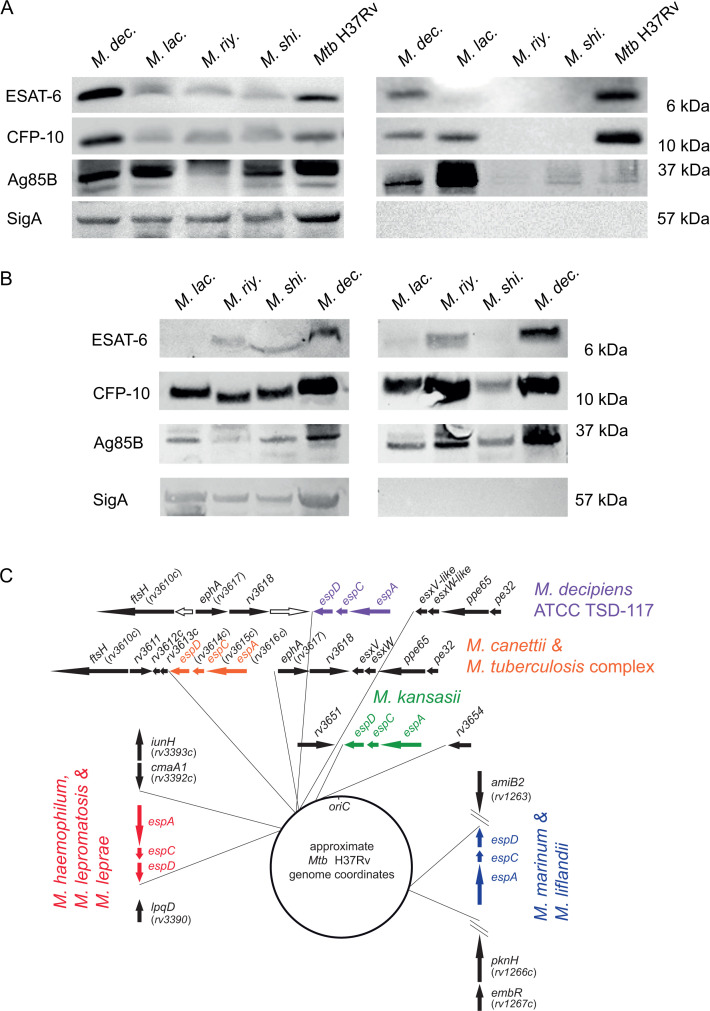
Fig 3.
Western blot-based evaluation of the presence of ESAT-6 and CFP-10 in whole-cell lysate (left panel) and supernatant (right panel) fractions of in vitro cultures of M. decipiens (M. dec.), M. lacus (M. lac.), M. riyadhense (M. riy.), M. shinjukuense (M. shi.), and Mtb H37Rv that were grown to an OD600nm of 0.6–0.8 using anti-ESAT-6, anti-CFP-10, as well as anti-Ag85B and anti-SigA control antibodies. An amount of 50 µg of proteins was migrated on SDS-PAGE for 30 min at 200 V and transferred onto a nitrocellulose membrane for 7 min at 110 V (A). Western blotting of whole-cell lysate (left panel) and supernatant (right panel) fractions from M. lacus (M. lac.), M. riyadhense (M. riy.), M. shinjukuense (M. shi.), and M. decipiens (M. dec.) cultures that were grown to an optical density higher than 1 (B). Genomic position of the orthologous genes of the espACD cluster that is present in the genome of M. decipiens, compared to the genomic locations of orthologous espACD clusters in selected other mycobacterial species that possess an espACD cluster in their genomes (C). Note that the orthologous flanking genes of the espACD cluster identified in the various species refer to the Mtb H37Rv gene nomenclature and relative genomic localization. Image adapted from reference (16) with permission and complemented with new information on the M. decipiens espACD cluster. Genes depicted as white arrows correspond to M. decipiens specific genes.

