
Figure 3.
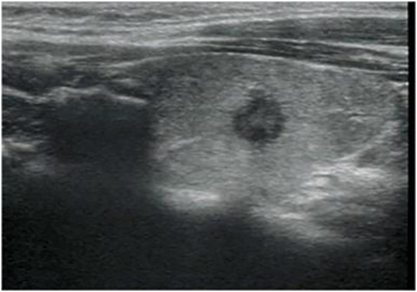
Longitudinal ultrasonography image from a 34-year-old woman with papillary thyroid carcinoma shows a 7.5-mm solid, hypoechoic, and irregular margin thyroid nodule with punctate echogenic foci, associated with no suspicious lymph node. The nodule was classified as EU-TIRADS 5:high risk or K-TIRADS 5:high suspicion. The nodular failed FNA based on nodule size.