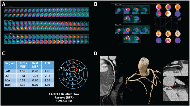
FIGURE 7. Coronary Microvascular Dysfunction in a Patient With Active Rheumatoid Arthritis Presenting With Dyspnea.
(A) 13N-ammonia Perfusion PET. Perfusion images demonstrate moderate ischemia in the left anterior descending (LAD) artery with vasodilator administration. (B) Functional imaging reveals normal LV volumes and systolic function at rest and stress (63% and 75%, respectively) without regional wall motion abnormalities. (C) Coronary flow reserve demonstrates globally reduced peak myocardial blood flow (normal >1.8) and associated reduction in coronary flow reserve (abnormal defined as CFR<2.0). The 17-segment model highlights the LAD relative flow reserve and a gradient suggestive of diffuse nonobstructive atherosclerosis. (D) Coronary CT was performed to rule out severe LAD stenosis which revealed evidence of a medium amount of predominantly noncalcified coronary plaque throughout the epicardial coronaries, resulting in minimal stenosis of the left main, proximal and mid-LAD, proximal left circumflex, and right coronary artery (shown is the LAD)

