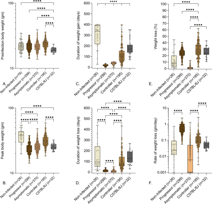
Fig 2.
Preinfection body weights and weight-related indicators of pulmonary TB in M. tuberculosis-infected mice. We infected 8- to 10-week-old, female Diversity Outbred mice and C57BL/6J mice with aerosolized M. tuberculosis bacilli and monitored as described in Materials and Methods. Non-infected, identically housed, and age- and gender-matched Diversity Outbred mice served as controls. Mice were euthanized at a predetermined timepoint, or if any one of three morbidity criteria developed: body condition score of <2, severe lethargy, or increased respiratory rate/effort. (A) Preinfection body weights, (B) peak body weight achieved during infection, (C) duration of weight gain, (D) duration of weight loss, (E) percentage of peak body weight lost, and (F) rate of weight lost are shown. Box-and-whiskers plots in all panels show interquartile range with whiskers at the minimum and maximum. Group names and sample sizes are shown on the X-axis. Statistical analyses were performed using Brown-Forsythe and Welch’s one-way analysis of variance followed by Dunnett’s T3 post-test. Non-significant P values are not shown. ****P < 0.0001.

