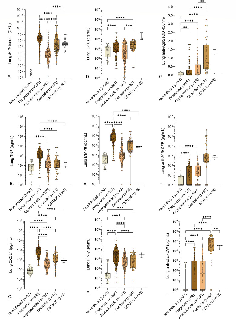
Fig 3.
Lung M. tuberculosis burden, cytokines, chemokines, and anti-M. tuberculosis antibodies in M. tuberculosis-infected mice. We infected 8- to 10-week-old, female Diversity Outbred mice and C57BL/6J mice with aerosolized M. tuberculosis bacilli and monitored as described in Materials and Methods. Non-infected, identically housed, and age- and gender-matched Diversity Outbred mice served as controls. Mice were euthanized at a predetermined timepoint or if any one of three morbidity criteria developed: body condition score of <2, severe lethargy, or increased respiratory rate/effort. We quantified M. tuberculosis colony-forming units in the lungs (A) and measured eight lung proteins using sandwich ELISAs (B–I). Box-and-whiskers plots in all panels show interquartile range with whiskers at the minimum and maximum. Sample sizes are shown in the X-axis. Statistical analyses were performed using Kruskal-Wallis one-way analysis of variance (ANOVA) with Dunn’s multiple comparisons post-tests (A) or Brown-Forsythe and Welch’s one-way ANOVA followed by Dunnett’s T3 post-test (B–F). Non-significant P values are not shown. **P < 0.01, ***P < 0.001, ****P < 0.0001.

