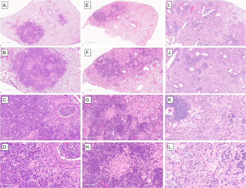
Fig 6.
Representative histopathological lesions in the lungs of M. tuberculosis-infected Diversity Outbred mice. We infected 8- to 10-week-old, female Diversity Outbred mice with M. tuberculosis bacilli by inhalation. Lung lobes were fixed, stained, and sectioned for microscopic examination. (A–D) Representative necrosuppurative lung lesions with bronchiolar obstruction in progressors. (E–H) Non-necrotizing lymphohistiocytic lung lesions in asymptomatic mice. (I–L) Diffuse, non-necrotizing lesions with abundant macrophages, foamy macrophages, scattered lymphocytic foci, and a few cholesterol clefts in controllers. Magnifications are ×2, ×4, ×20, and ×40.

