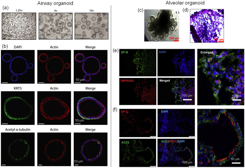
Figure 3.
Lung organoid cultures. Left column: airway organoid [van der Vaart 2021a]. (a) Brightfield images of airway organoids. (b) Confocal images showing Cytokeratin 5 (KRT5, basal cell marker) and Acetyl α-tubulin (ciliated cell marker) of the organoid. Right column: alveolar organoid [Tiwari 2021]. (c) Phase-contrast image of lung alveolar organoids. (d) Hematoxylin & eosin (H&E) staining of alveolar-like morphology. (e-f) Confocal images showing AT2 cells ((e) SP-B and (f) SP-C) co-labeled with (e) TMPRSS2 and (f) ACE2.

