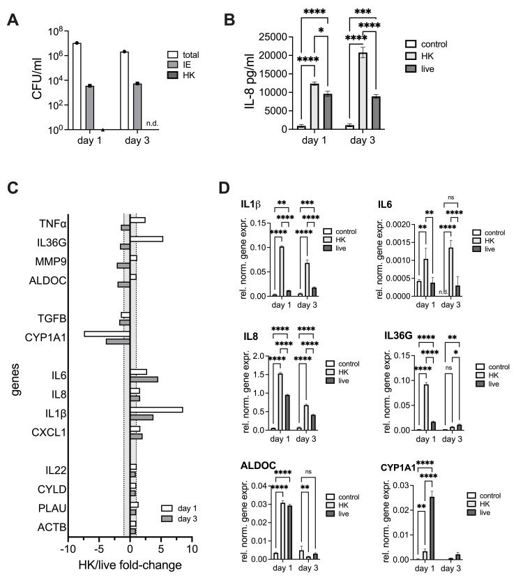
Fig 7. Infection of NHNE from a single donor with live or heatkilled (HK) NTHi.
Panel A: NTHi CFU/ml on different days of infection, n.d.–none detected, Panel B: IL-8 concentrations detected using ELISA in apical wash fluids for unifected NHNE and NHNE infected with heat-killed or live NTHi, Panel C: Gene expression differences between NHNE infected with heat-killed (HK) or live NTHi expressed as HK/live fold-changes. Grey shading–area where no change in gene expression occurred, Panel D: Gene expression in unifected NHNE and NHNE infected with heat-killed (HK) or live NTHi. Values were determined by qPCR and expression normalized to expression of ACTB. NHNE were infected with NTHI 86-028NP. Statistical testing used 2-Way ANOVA with Tukey’s multi-comparison correction.

