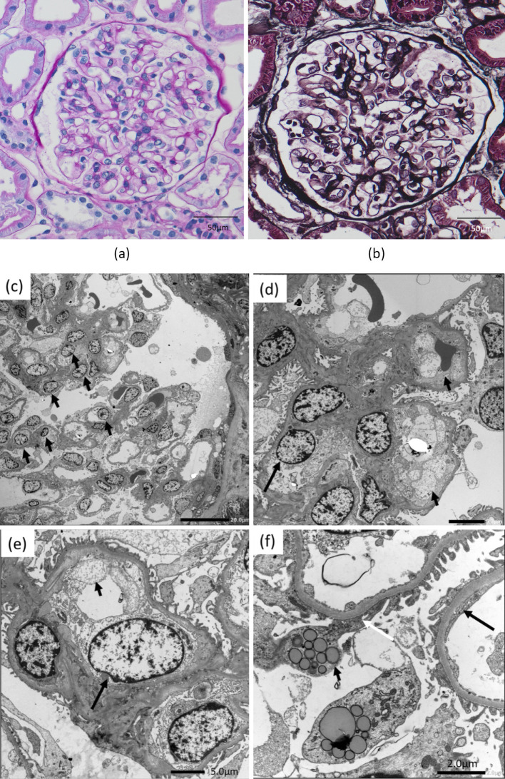
Figure 1.
Kidney biopsy findings. a+b: Light microscopy findings: Glomerular endothelial cell proliferation was slightly prominent. a: Periodic acid-Schiff (original magnification ×400; bar=50 μm). b: Periodic acid methenamine silver staining (original magnification, ×400; bar=50 μm). c-f: Electron microscopy findings. c: At low magnification, endothelial cell proliferation (arrow) was noted. d and e: Endothelial cell swelling (large arrow) and subendothelial edema (small arrow) were observed at high magnification. f: Endothelial fenestrations are not regularly arranged, and fenestration fusion (large arrow) is observed. Many areas of foot process effacement (white arrow) were seen. Vacuolar structures (small arrow) were present in the podocyte.

