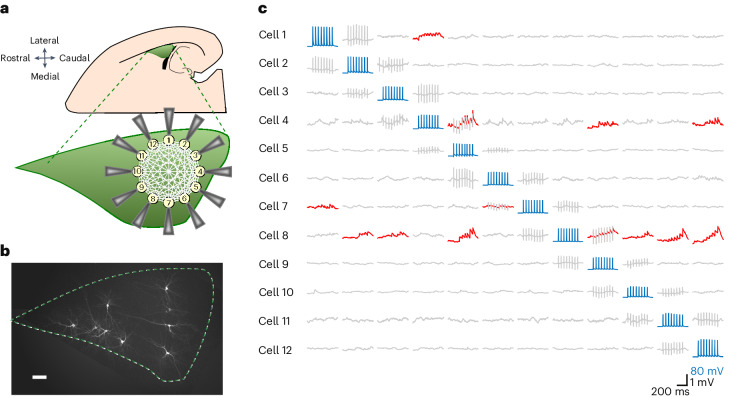
Fig. 1. Schematic view of 12 whole-cell patch-clamp recordings in acute horizontal brain slices and post hoc fluorescent biocytin staining.
a, The preparation involved half-hemisphere horizontal slices from a 2- to 3-week-old rat that contained the LA (green), which is easily recognizable by being bordered by the external capsule on the lateral border and the hippocampus and the lateral ventricle on the caudal border. After gaining whole-cell access to up to 12 neurons at a time, electrophysiological recordings were performed. b, Post hoc staining of ten biocytin-labeled neurons in the LA (n = 90 slices). Scale bar, 100 μm. c, A 20-Hz train of eight APs, in addition to a follow-up single AP (not shown), were elicited successively in each recorded neuron (blue traces) while recording spontaneous activity from the remaining neurons. Time-locked evoked responses (red traces) indicated a direct synaptic connection. Columns showing sequentially evoked APs (in blue) in cells 1–12 by injections of a 2 nA 3 ms–1 current and simultaneous membrane potentials (in gray) of nonstimulated cells. uEPSPs (in red) were averaged over 15 trials recorded in 14- to 19-day-old Wistar rats of both sexes.

