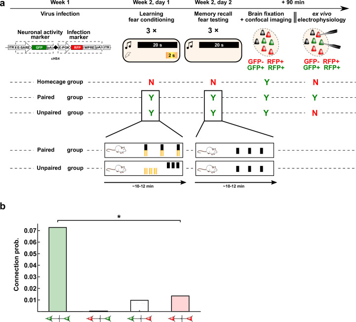
Extended Data Fig. 7. Detailed experimental outline of viral labeling of activated neurons and their connection probability following fear conditioning.
a, Protocol timeline: from virus infusion to fear conditioning and recall of the memory (testing), followed by imaging or electrophysiology 90 min later. Viral construct: bilaterally injected with glass pipettes targeting the LA. The viral construct is flanked by Inverted Terminal Repeats (ITR), expresses d2Venus under the enhanced synaptic activity response element (E-SARE) and the red fluorescent protein (RFP; FP635) under the constitutive enhanced phosphoglycerate kinase promoter (E-PGK). The woodchuck hepatitis post-transcriptional regulatory element (WPRE) enhances expression levels and is followed by a poly-adenylation (pA) signal. Behavior: Rats in the Paired group were fear conditioned by co-terminated pairing of the conditioned stimulus (CS, 20 second tone, black bars) and unconditioned stimulus (US, electric shock, yellow bars) thrice, at random intervals (from 60 s to 180 s – determined by Matlab’s rand function). The Unpaired group received 3xUS and 3xCS at the start and end of the conditioning session, respectively. Fear memory recall was tested by 3xCS presentations for both Paired and Unpaired groups. The home-cage group was not exposed to CS, US or conditioning context. Rats were sacrificed 90 min. after conditioning when GFP expression is optimal. Confocal imaging: GFP+ and RFP+ neurons were counted as memory-recall-participating and/or infected, respectively. Ex vivo electrophysiology (Paired group): multi-electrode whole-cell patch-clamp was performed on GFP+ and GFP− neurons to assess connectivity and connection strength of connections recruited during memory recall. b, Connectivity was significantly higher between (recruited) GFP+-GFP+ neurons (n = 15 slices with GFP+-GFP+ connections; 49 GFP+ neurons, with 6 out of 107 possible connections) than between GFP−-GFP− neurons (n = 39 slices with GFP−-GFP− connections; 137 GFP− neurons, with 5 out of 369 possible connections) (Two-sided Wilcoxon rank sum test with continuity correction, W = 378.5, P = 0.0292 (*)); however, overall connectivity calculated from all connections between recruited and non-recruited neurons was unchanged at ~2%, that is similar to connectivity in naïve homecage controls (see Fig. 3a, red-dashed line), suggesting that plasticity does not increase the total number of connections within the LA.

