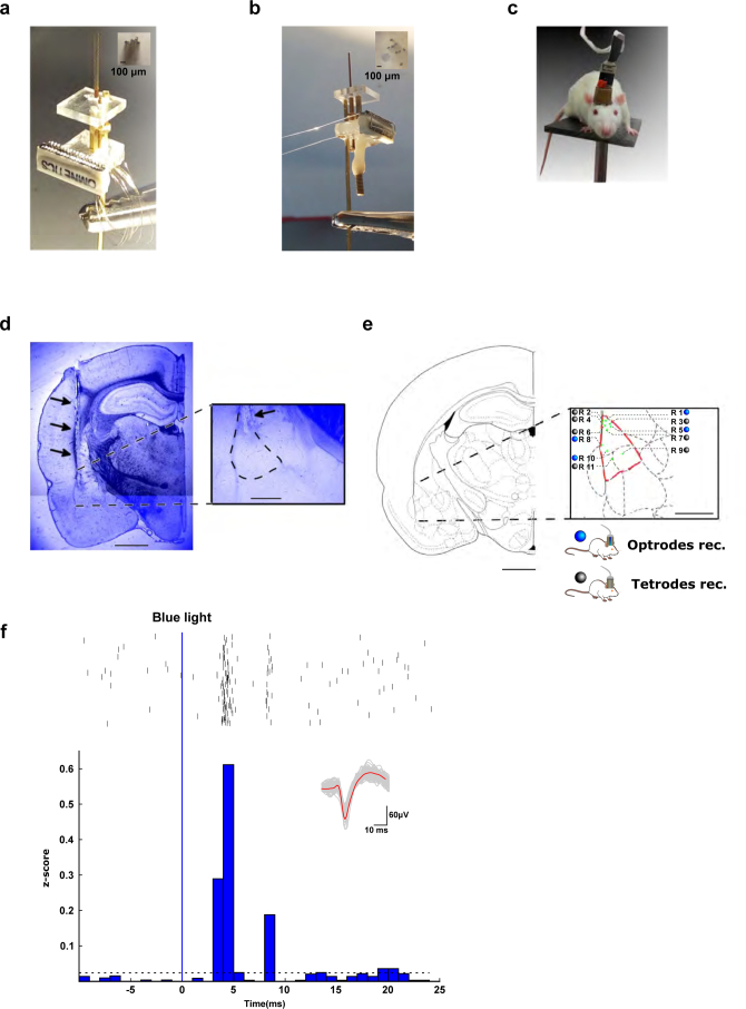
Extended Data Fig. 9. In vivo recordings – tetrode implantation.
a, Microdrive with 8 tetrodes (32 electrodes total) used for in vivo electrophysiological recordings in freely-moving rats. b, Optical fiber that allows – in addition to recording – stimulation with blue light. c, Rat implanted with a microdrive. d, DAPI staining of a coronal section of the lateral amygdala (inset: LA, dashed outline) showing the localization of the implanted tetrodes (black arrows). Image shows a representative example of tetrode or optrode placements in 11 rats. Scale bars indicate 2 mm or 0.5 mm (inset). e, Localization of all electrode tips, determined post-hoc. Scale bar indicates 1 mm. f, Example of Channelrhodopsin-2-expressing neuronal unit responding to a 1-ms blue-light pulse with time-locked spikes following the blue stimulus within a few ms (top, rasterplot data, n = 540 repetitions; bottom, z-score representation with the horizontal dashed line representing 3 standard deviations of baseline activity); inset: spike waveforms of recorded unit.

