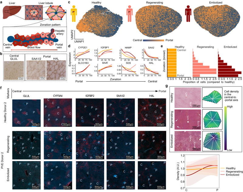
Fig. 3. Spatial zonation patterns are altered in regenerating and embolized tissue hepatocytes.
a Schematic illustrating the structure of a liver lobule. b 3,3′-Diaminobenzidine (DAB) stainings of central and portal zone-specific protein expression within healthy tissue hepatocytes. c Gene expression signature of zonation within healthy (right), regenerating (middle), and embolized (left) tissue hepatocytes. Cells in UMAP plots are colored based on the cumulative expression of central (blue) or portal (orange) zonation marker genes. d Pseudozonation expression patterns of representative zonation marker genes for each medical condition. e Histograms showing the proportion of hepatocytes in zonation bins across conditions. f Immunofluorescence tissue staining for liver tissue for various hepatocyte zonation markers. P portal vessel, C central vessel. See also Supplementary Figs. 9, 10. g, Top: Representative H/E stainings showing liver lobule segmentation to measure cell density along a central-to-portal axis; Bottom: Mean ± s.e. for cell density along the central-to-portal axis in each condition. Source data are provided as a Source Data file for (d, e).

