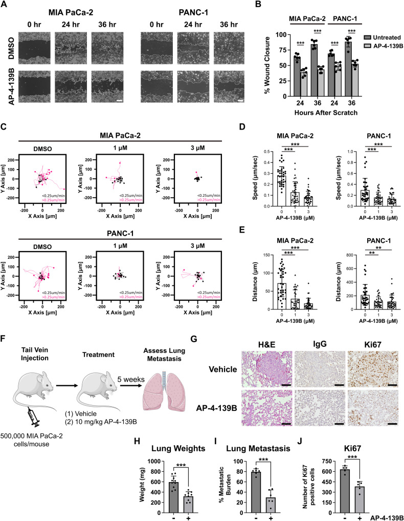
Fig. 4. HSP70 inhibition limits PDAC cell migration in vitro and metastasis in vivo.
A Primary (PANC-1) and metastatic (MIA PaCa-2) PDAC cells were seeded and allowed to form a confluent monolayer. Cells were scratched with a pipet tip and treated with DMSO or AP-4-139B and then imaged at 0, 24 and 36 h. Bar scale: 250 µm. B Quantification of the percentage of wound closure in (A) had 0, 24, and 36 h in DMSO versus AP-4-139B treated cells. The data depicted represent one representative containing data from three independent wells from a single experiment. n = 3 independent experiments; ***p < 0.001. C PANC-1 and MIA PaCa-2 cells were treated with the indicated doses of AP-4-139B for 24 h and were then analyzed for single-cell motility by time-lapse video-microscopy in 2D contour plots. The cutoff velocities for slow moving (black) or fast-moving (pink) cells are indicated. Quantification of the average speed of cell movements (D) and total distance traveled by individual cells (E). The mean ± SD speed of cell motility (µm/min), distance traveled (µm), and p values are indicated (n= approximately 35 cells per condition tested). **p < 0.01, ***p < 0.001. F Schematic representation of the metastasis assay. MIA PaCa-2 cells (5 × 10⁵) cells were injected into the tail vain of 8- to 10-week-old female NSG mice. Mice were treated with intraperitoneal (i.p.) injection of 10 mg/kg AP-4-139B every 48 h. After 5 weeks, the lungs of mice were formalin-fixed, and H&E stained and assessed for the presence of metastatic nodules. G Representative H&E and Ki67 images of lung metastases from NSG mice injected with MIA PaCa-2 cells in the tail vein, followed by treatment with vehicle or AP-4-139B. Bar scale: 100 µm. Rabbit IgG was used as a negative control for IHC analysis. Quantification of lung weights, metastatic burden, and Ki67 staining. Quantification of (H) was performed on all mice in the study, while quantification of (I, J) was performed on n = 5-6 mice per treatment group.

