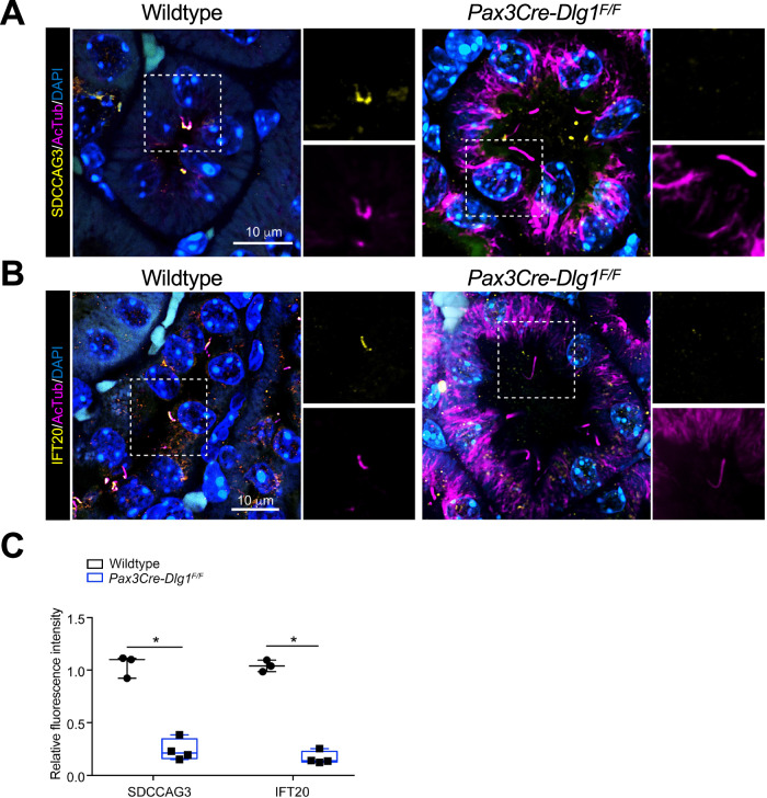
Figure 4. Conditional loss of DLG1 in mouse kidney leads to impaired ciliary localization of SDCCAG3 and IFT20.
(A, B) Immunofluorescence staining of SDCCAG3 (A) or IFT20 (B), both in yellow, and acetylated α-tubulin (AcTub, magenta) in kidney sections from wild-type and Pax3Cre-Dlg1F/F mice. (C) Quantification of relative MFI of SDCCAG3 and IFT20 in cilia of wild-type (n = 3) and Pax3Cre-Dlg1F/F (n = 4) mice, respectively. The levels from control mice were set to 1, and the ciliary levels from mutant mice were compared to that (i.e., relative fluorescence intensity). Data shown are the average values from each mouse. The vertical segments in box plots show the first quartile, median, and third quartile. The whiskers on both ends represent the maximum and minimum values for each dataset analyzed. Statistical analysis was performed two-tail unpaired t-test. *P < 0.05, **P < 0.01. Source data are available online for this figure.

