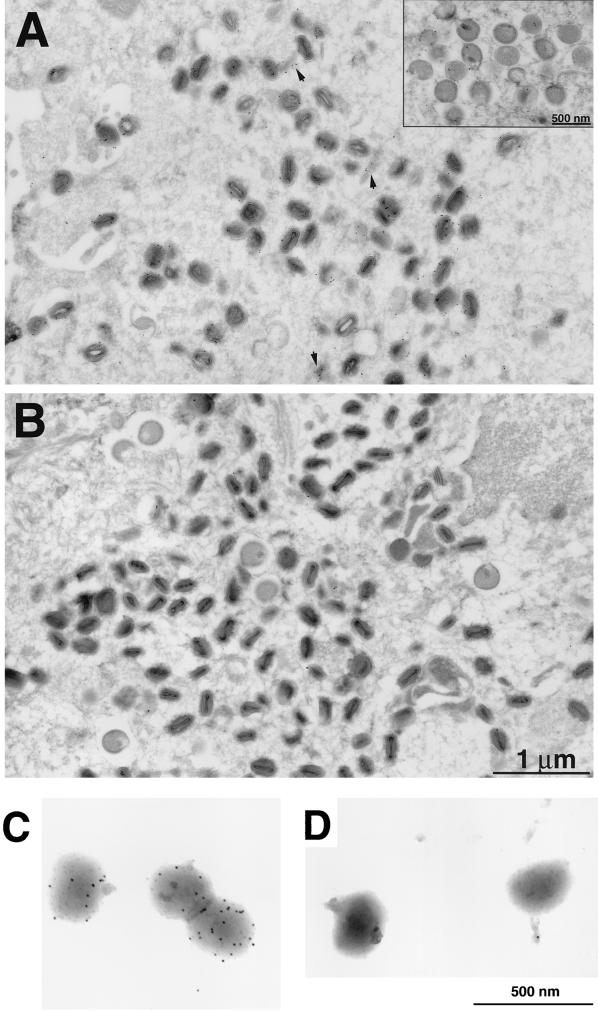
FIG. 5.
Localization of the A9L-HA protein by immunoelectron microscopy. BS-C-1 cells were infected with either vA9L-HA (A) or vT7lacOI (B) for 22 h, fixed in paraformaldehyde, cryosectioned, and incubated with MAb HA.11 followed by rabbit anti-mouse IgG and then protein A conjugated to 10-nm-diameter colloidal gold. Electron micrographs of these samples are shown with a 1-μm and a 500-nm marker (inset). Arrowheads point to unidentified structures with associated gold grains. Grids containing purified intact vA9L-HA (C) or WR (D) virions were stained as in panels B and C.

