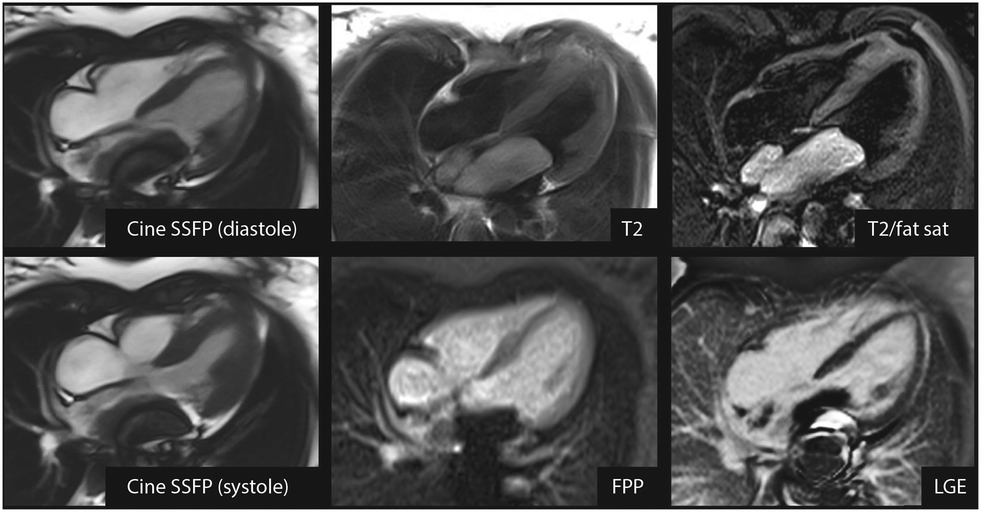
FIGURE 5.

High-Grade Leiomyosarcoma (Malignant Tumor) Incorrectly Diagnosed as a Myxoma
CMR of a 13-year-old child with palpitations and a large mass attached to the posterior wall of the left atrium, prolapsing through the mitral valve in diastole. Metallic susceptibility artifact obscures a portion of the mass. The mass is hyperintense on T2-weighted imaging with and without fat suppression, does not enhance on FPP imaging, and does not enhance on LGE imaging. Reviewer 1 and the clinical CMR report indicated myxoma as a single diagnosis, whereas reviewer 2 indicated a differential diagnosis of myxoma, thrombus, and malignant tumor. FPP = first-pass perfusion; LGE = late gadolinium enhancement; SSFP = steady-state free precession; T2/fat sat = T2-weighted imaging with fat suppression; other abbreviation as in Figure 2.
