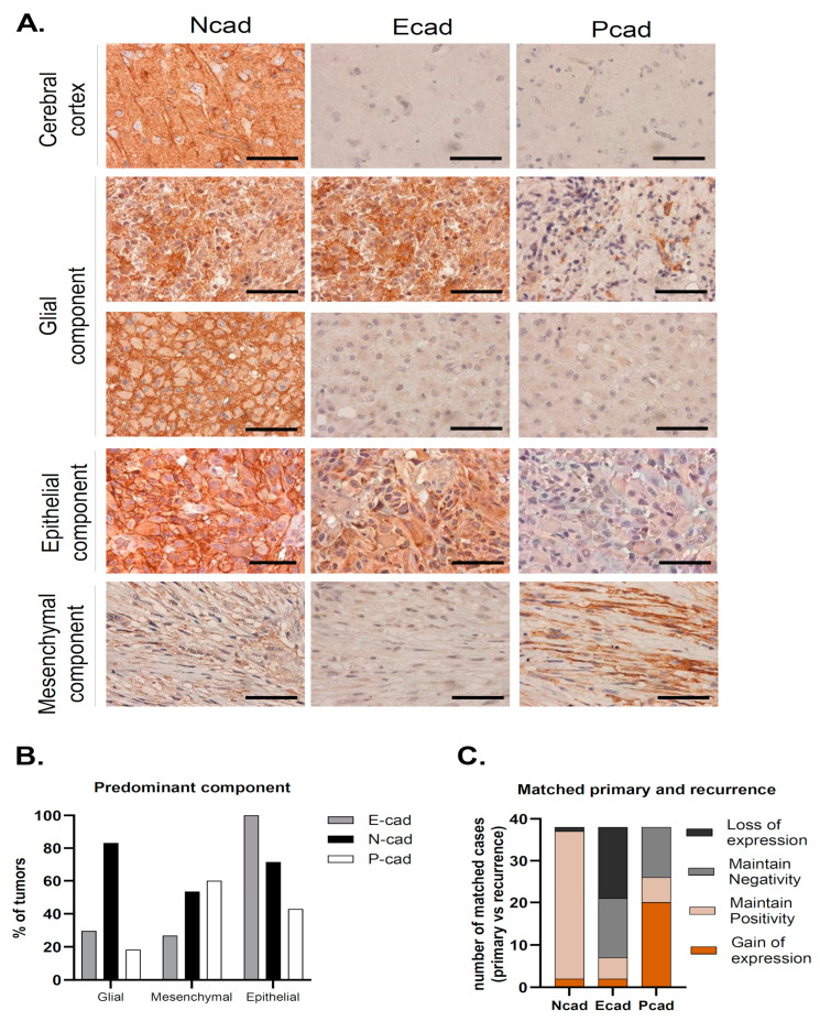
Figure 1.
N-, E- and P-cadherin expression in Glioblastoma. (A) Representative cases of predominant glial, epithelial and mesenchymal component tumors. In tumors with a predominant glial component, N-cadherin positivity was observed in the majority of the cases and was frequently strong, with membrane staining. In some cases, E-cadherin positivity was also observed. Tumors with a predominant epithelial component stained for E-cadherin in all cases, which was frequently accompanied by N-cadherin staining, as observed in this case. Finally, tumors with a predominant mesenchymal component most commonly stained for both N-cadherin and P-cadherin (scale bar corresponds to 50 μm). (B) N-cadherin expression was high in all tumor subtypes. In tumors with a predominant mesenchymal component, P-cadherin and N-cadherin expression was observed in the vast majority of cases. Strikingly, all cases with a predominant epithelial component were positive for E-cadherin. (C) Upon tumor recurrence, 97.4% of the tumors were positive for N-cadherin (maintain positive + gain of positivity). Gain of P-cadherin expression was the most common event which was observed in over half of the cases (51.3%). This leads to an important increase in P-cadherin positivity in recurrence when compared to the primary tumor (20.8% vs. 65.8%). On the contrary, E-cadherin positivity is lower in recurrence (31% vs. 20.8%), due to E-cadherin loss of staining in matched primary-recurrent cases (43.6%).

