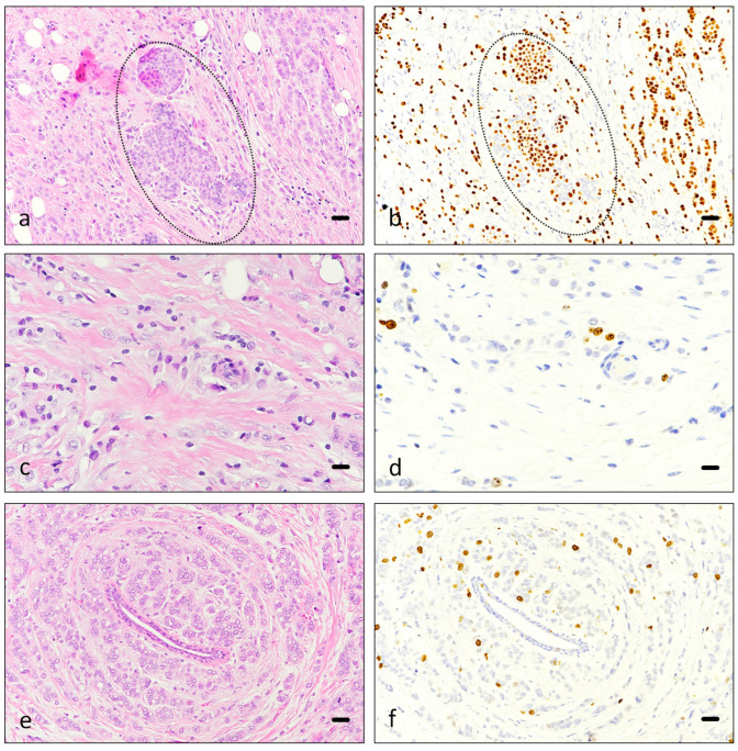
Figure 4.
Ki67 immunostaining. Hematoxylin-eosin (a,c,e) and Ki67 immunostaining (b,d,f) of breast carcinoma, corresponding to consecutive slides ((a,b), (c,d) and (e,f)). Images (a,b) correspond to an infiltrating lobular carcinoma, including in situ carcinoma component (discontinuous line). Images (c,d) and (e,f) depict ductal carcinoma in two different patients. A benign duct is centrally located in images (e,f). Scale bar 50 µm (a–f).

